Comparative Study
. 2004 Mar;72(3):1760-6.
doi: 10.1128/IAI.72.3.1760-1766.2004.

Fusarium oxysporum as a multihost model for the genetic dissection of fungal virulence in plants and mammals

Affiliations
Comparative Study

Fusarium oxysporum as a multihost model for the genetic dissection of fungal virulence in plants and mammals

Montserrat Ortoneda et al. Infect Immun. 2004 Mar.

Abstract

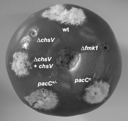
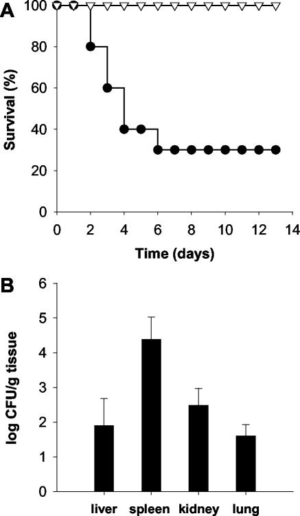
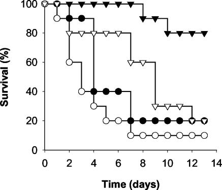
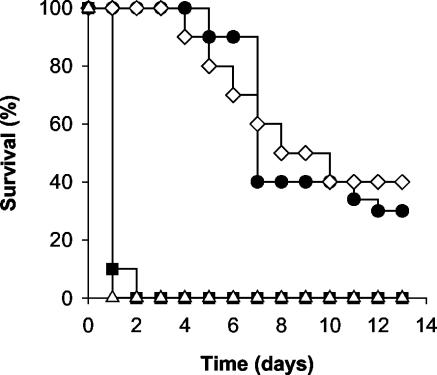
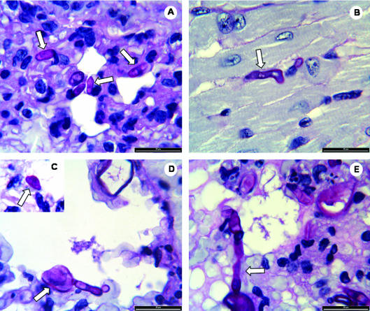
Fungal pathogens cause disease in plant and animal hosts. The extent to which infection mechanisms are conserved between both classes of hosts is unknown. We present a dual plant-animal infection system based on a single strain of Fusarium oxysporum, the causal agent of vascular wilt disease in plants and an emerging opportunistic human pathogen. Injection of microconidia of a well-characterized tomato pathogenic isolate (isolate 4287) into the lateral tail vein of immunodepressed mice resulted in disseminated infection of multiple organs and death of the animals. Knockout mutants in genes encoding a mitogen-activated protein kinase, a pH response transcription factor, or a class V chitin synthase previously shown to be implicated in virulence on tomato plants were tested in the mouse model. The results indicate that some of these virulence factors play functionally distinct roles during the infection of tomato plants and mice. Thus, a single F. oxysporum strain can be used to study fungal virulence mechanisms in plant and mammalian pathogenesis.


Figures

FIG. 1.
FIG. 1.
Invasive growth of F. oxysporum strains on tomato fruits. Fruits were inoculated by injecting 5 × 10⁵ microconidia of the following strains: wild-type strain 4287, MAPK mutant Δfmk1, pacC loss-of-function mutant pacC+/−, dominant-activating mutant pacCc, chitin synthase mutant ΔchsV complemented with the wild-type chsV allele, and chitin synthase mutant ΔchsV. The photograph was taken after 5 days of incubation at 100% relative humidity.
FIG. 2.
FIG. 2.
F. oxysporum strain 4287 causes systemic infection and death in immunodepressed mice. (A) Groups of 10 immunodepressed mice were infected with 2 × 10⁷ living (•) or heat-killed (▿) microconidia by lateral tail vein injection. The percent survival was plotted for 13 days. The data shown are from one representative experiment. (B) Four surviving mice were sacrificed on day 13 postinfection, and homogenates from the indicated organs were quantitatively cultured on PDA medium.
FIG. 3.
FIG. 3.
Virulence of gene knockout mutants of F. oxysporum on immunodepressed mice. Groups of 10 immunodepressed mice were infected with 2 × 10⁷ microconidia of the wild-type strain 4287 (•), MAPK mutant Δfmk1 (○), loss-of-function mutant pacC+/− (▾), or dominant-activating mutant pacCc (▿). The percent survival was plotted for 13 days. The data shown are from one representative experiment.
FIG. 4.
FIG. 4.
F. oxysporum mutants lacking the chitin synthase ChsV cause “fast killing” on mice. (A) Groups of 10 immunodepressed mice were infected with 2 × 10⁷ microconidia of the wild-type strain 4287 (•), chitin synthase mutant ΔchsV (▪), heat-killed conidia of the ΔchsV mutant (▵), or a ΔchsV mutant complemented with a wild-type chsV allele (⋄). The percent survival was plotted for 13 days. The data shown are from one representative experiment.
FIG. 5.
FIG. 5.
Histological analysis of mice infected with F. oxysporum strains. Sections of embedded tissues from different organs (A and C to E, lung; B, heart) of mice inoculated with microconidia of F. oxysporum wild-type strain (A and B) or chitin synthase mutant ΔchsV (C to E) and sacrificed 24 h after inoculation were stained with hematoxylin-eosin, periodic acid-Schiff, and methenamine silver and then examined by light microscopy. (A) Characteristic ellipsoidal microconidia of wild-type strain 4287 in the interstitial space; (B) germinated microconidium of wild-type strain 4287; (C) lemon-shaped microconidium of the ΔchsV mutant; (D) swollen ungerminated and germinated microconidia of the ΔchsV mutant; (E) germinated conidia of the ΔchsV mutant growing in the interstitial spaces and provoking an edema. Bar, 20 μm.

Similar articles

Cited by

References

    1. Beckman, C. H. 1987. The nature of wilt diseases of plants. American Phytopathological Society, St. Paul, Minn.
    1. Boutati, E. I., and E. J. Anaissie. 1997. Fusarium, a significant emerging pathogen in patients with hematologic malignancy: ten years' experience at a cancer center and implications for management. Blood 90:999-1008. - PubMed
    1. Buer, J., and R. Balling. 2003. Mice, microbes and models of infection. Nat. Rev. Genet. 4:195-205. - PubMed
    1. Cabib, E., D. H. Roh, M. Schmidt, L. B. Crotti, and A. Varma. 2001. The yeast cell wall and septum as paradigms of cell growth and morphogenesis. J. Biol. Chem. 276:19679-19682. - PubMed
    1. Caracuel, Z., M. I. Roncero, E. A. Espeso, C. I. Gonzalez-Verdejo, F. I. Garcia-Maceira, and A. Di Pietro. 2003. The pH signalling transcription factor PacC controls virulence in the plant pathogen Fusarium oxysporum. Mol. Microbiol. 48:765-779. - PubMed

Publication types

Substances