. 2004 Mar;15(3):663-73.
doi: 10.1097/01.asn.0000113553.62380.f5.

Mercuric conjugates of cysteine are transported by the amino acid transporter system b(0,+): implications of molecular mimicry

Affiliations

Mercuric conjugates of cysteine are transported by the amino acid transporter system b(0,+): implications of molecular mimicry

Christy C Bridges et al. J Am Soc Nephrol. 2004 Mar.

Abstract

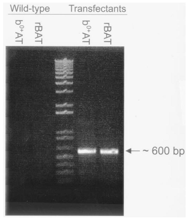
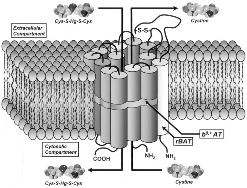
Humans and other mammals continue to be exposed to various forms of mercury in the environment. The kidneys, specifically the epithelial cells lining the proximal tubules, are the primary targets where mercuric ions accumulate and exert their toxic effects. Although the actual mechanisms involved in the transport of mercuric ions along the proximal tubule have not been defined, current evidence implicates mercuric conjugates of cysteine, primarily 2-amino-3-(2-amino-2-carboxyethylsulfanylmercuricsulfanyl)propionic acid (Cys-S-Hg-S-Cys), as the most likely transportable species of inorganic mercury (Hg(2+)). Because Cys-S-Hg-S-Cys and the amino acid cystine (Cys-S-S-Cys) are structurally similar, it was hypothesized that Cys-S-Hg-S-Cys might act as a molecular mimic of cystine at one or more of the amino acid transporters involved in the luminal absorption of this amino acid. One such candidate is the Na(+)-independent heterodimeric transporter system b(0,+). Therefore, the transport of Cys-S-Hg-S-Cys and cystine was studied in MDCK II cells that were or were not stably transfected with b(0,+)AT-rBAT. Transport of Cys-S-Hg-S-Cys and cystine across the luminal plasma membrane was similar in the transfected cells, indicating that Cys-S-Hg-S-Cys can behave as a molecular mimic of cystine at the site of system b(0,+). Moreover, only the b(0,+)AT-rBAT transfectants became selectively intoxicated during exposure to Cys-S-Hg-S-Cys. These findings indicate that system b(0,+) likely contributes to the nephropathy induced by Hg(2+) in vivo. These data represent the first direct molecular evidence for the participation of a specific transporter in the luminal uptake of a large divalent metal cation in proximal tubular cells.


Figures

Figure 1
Figure 1
Agarose gel showing reverse transcription-PCR analysis of the steady-state expression of mRNA encoding mouse b0,+AT and human rBAT in wild-type and b0,+AT-rBAT-transfected MDCK II cells. The expected sizes of the reverse transcription-PCR products, as predicted from the positions of the primers, were 589 bp for mouse b0,+AT and 600 bp for human rBAT. Transcripts for mouse b0,+AT and human rBAT were not detected in wild-type cells.
Figure 2
Figure 2
Laser scanning confocal microscopic images of b0,+AT-rBAT-transfected MDCK II cells labeled with antibodies to mouse b0,+AT and human rBAT, followed by incubation with Texas Red-labeled and FITC-labeled secondary antibodies, respectively. Upper panels, images of cells in a horizontal (xy) plane; lower panels, images of cells in a vertical (zy) plane. (A) Colocalization of mouse b0,+AT and human rBAT on the plasma membranes of transfected cells. The distribution of mouse b0,+AT is represented by red fluorescence, and the distribution of human rBAT is represented by green fluorescence. The yellow fluorescence represents the colocalization of these two proteins. The image in the vertical plane demonstrates the colocalization of these proteins on the apical plasma membrane of these cells. (B) Control experiment in which the primary antibody against human rBAT was omitted. Scale bars = 5 μm.
Figure 3
Figure 3
Time course of uptake of 5 μM cystine (containing [35S]cystine) (A) or 5 μM inorganic mercury, as the mercuric conjugate of cysteine, 2-amino-3-(2-amino-2-carboxyethylsulfanylmercuricsulfanyl)propionic acid (Cys-S-Hg-S-Cys) (containing 203Hg2+) (B), in wild-type and b0,+AT-rBAT-transfected cells. Uptake was performed at 37°C for times ranging from 5 to 90 min. Samples were obtained for estimation at the indicated times. Results are presented as means ± SEM. Data represent three experiments performed in quadruplicate. *, significantly different (P < 0.05) from the mean for the corresponding group of wild-type cells; +, significantly different from the mean for the same cell type at the initial time point; ++, significantly different from the mean for the same cell type at each of the two preceding time points; +++, significantly different from the mean for the same cell type at each of the three preceding time points.
Figure 4
Figure 4
Saturation kinetics for the transport of cystine or inorganic mercury (Hg2+), as the mercuric conjugate of cysteine, Cys-S-Hg-S-Cys, in wild-type and b0,+AT-rBAT-transfected MDCK II cells. Cells were incubated for 30 min at 37°C with either 5 μM cystine (containing [35S]cystine) (A) or 5 μM Cys-S-Hg-S-Cys (containing 203Hg2+) (B), in the presence of unlabeled cystine (25 to 750 μM) or Cys-S-Hg-S-Cys (1 to 100 μM), respectively. Results are presented as means ± SEM. Data represent three experiments performed in quadruplicate. *, significantly different (P < 0.05) from the mean for the corresponding group of wild-type cells; +, significantly different from the mean for the same cell type at the initial concentration; ++, significantly different from the mean for the same cell type at each of the preceding concentrations; +++, significantly different from the mean for the same cell type at each of the preceding concentrations; ++++, significantly different from the mean for the same cell type at each of the preceding concentrations.
Figure 5
Figure 5
Temperature dependence of uptake of 5 μM cystine (containing [35S]cystine) (A) or 5 μM inorganic mercury, as the mercuric conjugate of cysteine, Cys-S-Hg-S-Cys (containing 203Hg2+) (B), in wild-type and b0,+AT-rBAT-transfected cells. Uptake was measured in the presence of unlabeled cystine (25 to 500 μM) or Cys-S-Hg-S-Cys (1 to 100 μM), respectively, at 37°C or 4°C. Results are presented as means ± SEM. Data represent three experiments performed in quadruplicate. *, significantly different (P < 0.05) from the mean for the corresponding groups of wild-type cells at 4°C and 37°C and significantly different from the mean for the corresponding group of transfected cells at 4°C; +, significantly different from the mean for the transfectants at the initial concentration at 37°C; ++, significantly different from the mean for the transfectants at each of the preceding concentrations at 37°C; +++, significantly different from the mean for the transfectants at each of the preceding concentrations at 37°C; ++++, significantly different from the mean for the transfectants at each of the preceding concentrations at 37°C.
Figure 6
Figure 6
Substrate specificity analyses of the uptake of cystine (containing [35S]cystine) (A) or inorganic mercury (Hg2+), as the conjugate of cysteine, Cys-S-Hg-S-Cys (containing 203Hg2+) (B), in wild-type and b0,+AT-rBAT-transfected MDCK II cells. Cells were incubated for 30 min at 37°C with either 5 μM cystine (containing [35S]cystine) or 5 μM Cys-S-Hg-S-Cys (containing 203Hg2+), in the presence of various unlabeled amino acids (at 3 mM, except for unlabeled cystine at 1 mM). Results are presented as means ± SEM. Data represent three experiments performed in quadruplicate. *, significantly different (P < 0.05) from the mean for the corresponding group of wild-type cells; +, significantly different from the mean for the control group of the corresponding cell type.
Figure 7
Figure 7
Uptake of 5 μM cystine (containing [35S]cystine) (A) or 5 μM inorganic mercury, as the mercuric conjugate of cysteine, Cys-S-Hg-S-Cys (containing 203Hg2+) (B), in the presence of unlabeled Cys-S-Hg-S-Cys (1 to 100 μM) or unlabeled cystine (25 to 1000 μM), respectively, in wild-type and b0,+AT-rBAT-transfected MDCK II cells. Cells were incubated for 30 min at 37°C. Results are presented as means ± SEM. Data represent three experiments performed in quadruplicate. *, significantly different (P < 0.05) from the mean for the corresponding group of wild-type cells; +, significantly different from the mean for the same cell type at the initial concentration; ++, significantly different from the mean for the same cell type at each of the preceding concentrations; +++, significantly different from the mean for the same cell type at each of the preceding concentrations; +++, significantly different from the mean for the same cell type at each of the preceding concentrations; ++++, significantly different from the mean for the same cell type at each of the preceding concentrations.
Figure 8
Figure 8
Efflux of 35S (A) and cellular contents of 35S after efflux (B) in wild-type MDCK II cells and b0,+AT-rBAT transfectants after exposure to cystine (containing 35S). Cells were exposed to 1 mM cystine for 10 min at 37°C and then incubated with buffer only or 1 mM unlabeled cystine, the mercuric conjugate of cysteine (Cys-S-Hg-S-Cys), arginine, or glutamate for 1 min at 37°C. Results are presented as means ± SEM. Data represent two experiments performed in duplicate. *, significantly different (P < 0.05) from the mean for the corresponding group of wild-type cells;+, significantly different from the mean for the group of transfectants exposed to buffer or glutamate; ++, significantly different from the mean for the transfectants exposed to buffer, cystine, arginine, or glutamate.
Figure 9
Figure 9
Uptake of inorganic mercury (Hg2+) in wild-type and b0,+AT-rBAT-transfected MDCK II cells exposed to 5 μM Hg2+ (containing 203Hg2+) and 20 μM cysteine (Cys), N-acetylcysteine (NAC), cysteinylglycine (CysGly), or GSH for 30 min at 37°C. Results are presented as means ± SEM. Data represent three experiments performed in quadruplicate. *, significantly different (P < 0.05) from the mean for the corresponding group of wild-type cells; +, significantly different from the mean for the group of transfected cells treated with Cys-S-Hg-S-Cys.
Figure 10
Figure 10
Cellular viability of wild-type cells and b0,+AT-rBAT transfectants after 24 h of exposure to various concentrations of Hg2+, in the form of the mercuric conjugate of cysteine (Cys-S-Hg-S-Cys) (A), the mercuric conjugate of GSH (GSH-S-Hg-S-GSH) (B), or HgCl2 (C). Results are presented as percent survival. Data represent two experiments performed in duplicate. *, significantly different (P < 0.05) from the mean for the corresponding group of wild-type cells; +, significantly different from the mean for the same cell type at the initial concentration; +++, significantly different from the mean for the same cell type at each of the preceding concentrations; +++, significantly different from the mean for the same cell type at each of the preceding concentrations; ++++, significantly different from the mean for the same cell type at each of the preceding concentrations.
Figure 11
Figure 11
Diagrammatic representation of the transport of cystine and the mercuric conjugate of cysteine (Cys-S-Hg-S-Cys) via system b0,+. The form of Hg2+ that is most likely presented to the luminal membrane of proximal tubular epithelial cells in vivo is a conjugate of cysteine, primarily in the form of Cys-S-Hg-S-Cys. As shown in this model, the molecular structure of Cys-S-Hg-S-Cys is very similar to that of the amino acid cystine (Cys-S-S-Cys). Given the structural similarity of cystine and Cys-S-Hg-S-Cys, it is probable that a transport system with a high affinity for cystine mediates the uptake of both compounds. We postulate that a likely candidate for this uptake is system b0,+. This carrier is an amino acid exchanger that mediates the transport of cystine, as well as a variety of neutral and cationic amino acids. As a heterodimeric transporter, it is composed of two subunits, b0,+AT (shaded cylinders) and rBAT (white cylinder). The linkage of these two subunits, via a disulfide bond (S-S), is essential for the formation of a functional transporter unit. Our data indicate that Cys-S-Hg-S-Cys is a transportable substrate of system b0,+. The data also indicate that Cys-S-Hg-S-Cys stimulates the efflux of cystine in b0,+AT-rBAT-transfected MDCK II cells. Because this efflux is specific to system b0,+, it likely that this transporter mediates the exchange of Cys-S-Hg-S-Cys and cystine.

Similar articles

Cited by

References

    1. Zalups RK, Lash LH. Advances in understanding the renal transport and toxicity of mercury. J Toxicol Environ Health. 1994;42:1–44. - PubMed
    1. Zalups RK. Molecular interactions with mercury in the kidney. Pharmacol Rev. 2000;52:113–143. - PubMed
    1. Berndt WO, Baggett JMcC, Blacker A, Houser M. Renal glutathione and mercury uptake by kidney. Fundam Appl Toxicol. 1985;5:832–839. - PubMed
    1. Tanaka T, Naganuma A, Imura N. Role of γ-glutamyltranspeptidase in renal uptake and toxicity of inorganic mercury in mice. Toxicology. 1990;60:187–198. - PubMed
    1. Tanaka-Kagawa T, Naganuma A, Imura N. Tubular secretion and reabsorption of mercury compounds in mouse kidney. J Pharmacol Exp Ther. 1993;264:776–782. - PubMed

Publication types