. 2004 May 15;557(Pt 1):175-90.
doi: 10.1113/jphysiol.2003.055772. Epub 2004 Feb 20.

Effects of uniform extracellular DC electric fields on excitability in rat hippocampal slices in vitro

Affiliations

Effects of uniform extracellular DC electric fields on excitability in rat hippocampal slices in vitro

Marom Bikson et al. J Physiol. .

Abstract

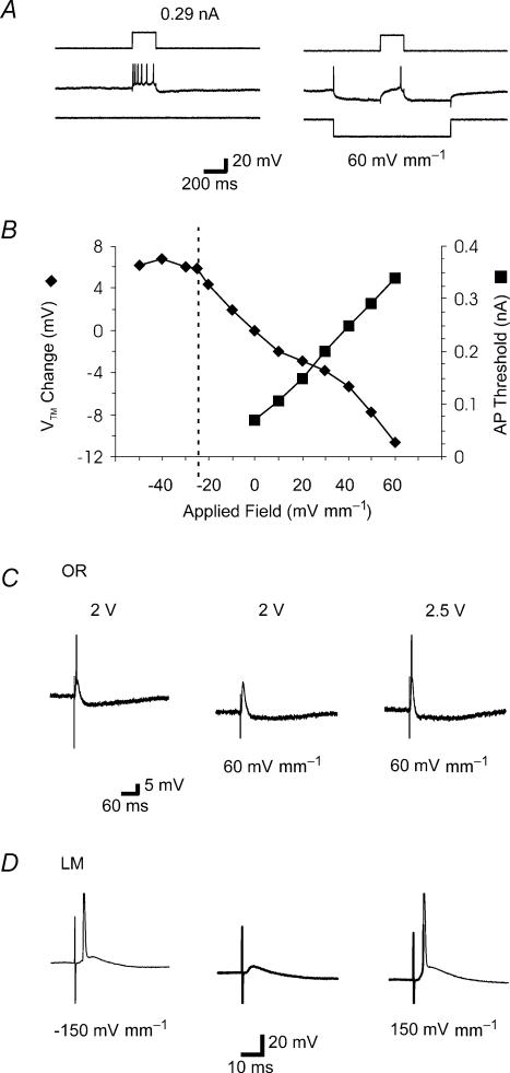
The effects of uniform steady state (DC) extracellular electric fields on neuronal excitability were characterized in rat hippocampal slices using field, intracellular and voltage-sensitive dye recordings. Small electric fields (</40/ mV mm(-1)), applied parallel to the somato-dendritic axis, induced polarization of CA1 pyramidal cells; the relationship between applied field and induced polarization was linear (0.12 +/- 0.05 mV per mV mm(-1) average sensitivity at the soma). The peak amplitude and time constant (15-70 ms) of membrane polarization varied along the axis of neurons with the maximal polarization observed at the tips of basal and apical dendrites. The polarization was biphasic in the mid-apical dendrites; there was a time-dependent shift in the polarity reversal site. DC fields altered the thresholds of action potentials evoked by orthodromic stimulation, and shifted their initiation site along the apical dendrites. Large electric fields could trigger neuronal firing and epileptiform activity, and induce long-term (>1 s) changes in neuronal excitability. Electric fields perpendicular to the apical-dendritic axis did not induce somatic polarization, but did modulate orthodromic responses, indicating an effect on afferents. These results demonstrate that DC fields can modulate neuronal excitability in a time-dependent manner, with no clear threshold, as a result of interactions between neuronal compartments, the non-linear properties of the cell membrane, and effects on afferents.


Figures

Figure 1
Figure 1. Schematic of electrophysiology and optical mapping experimental design
In all studies, uniform electric fields were generated by passing current between two large parallel silver—silver chloride wires positioned in the bath across the slice. A, for electrophysiological measurements activity was monitored in the CA1 pyramidal cell layer with a glass microelectrode. In some experiments, an additional field electrode was positioned in an iso-potential (see text) to remove the uniform field artefact. In some experiments, activity was evoked with a bipolar nichrome stimulating electrode positioned in either stratum lacunosum moleculare (LM) or stratum oriens (OR). (Not to scale.) B, schematic drawing of the optical apparatus for fluorescence measurement and the rat hippocampal slice preparation. The microscope objective forms a real magnified image of the preparation at the microscope image plane. A 16 × 16 array of photodetectors, positioned at the image plane, records the changes in light intensities that are related to neuronal activity. Epi-illumination with a 20 ×, 0.5 NA, long working distance, objective was used. The preparation was illuminated with the green portion (EX: excitation filter 520 ± 45 nm) of the output of a halogen lamp. Fluorescence emission from the preparation was selected by means of a dichroic mirror (DM: 570 nm) and an emission filter (EM: 590 nm). The output of each detector was individually amplified, multiplexed, digitized and stored in a PC.
Figure 2
Figure 2. Effect of applied uniform electric fields on population spikes evoked by oriens stimulation
A, top, stimulus protocol. Population spikes were evoked continuously at 0.5 Hz. One-second electric fields were applied 500 ms before the orthodromic pulse. Bottom, population spikes evoked before, during and after application of 40 mV mm−1 uniform electric fields. Pre- and post-traces are overlaid. In this and subsequent figures, the orthodromic stimulation artefact is clipped. B, effect of varied amplitude electric fields on population spike amplitude and delay; summary of single slice.
Figure 3
Figure 3. Effect of ±60 mV mm−1 applied electric fields on population spikes evoked by varying intensity oriens stimulation
Top to bottom, orthodromic stimulus intensity was incrementally increased. At each orthodromic stimulus intensity, the effect of −60 mV mm−1 (middle) and +60 mV mm−1 (right) electric fields was determined. Note that no spontaneous activity was observed in this slice at these field strengths.
Figure 4
Figure 4. Effect of ±100 mV mm−1 applied electric fields on evoked population spikes and spontaneous activity
A, top, no spontaneous activity was observed in the absence of applied electric fields or after application of +100 mV mm−1 electric fields. Middle, −100 mV mm−1 fields (1.0 second) induced spontaneous epileptiform activity. Population spikes were evoked by oriens stimulation continuously at 0.5 Hz. Bottom, expansion of field traces before (left), during (middle), and after (right) application of −100 mV mm−1 electric fields. Note that postfield evoked response (right) did not return to control levels (left). The orthodromic stimulus artefact was removed in the expansion insets. B, effect of prolonged application of −100 mV mm−1 electric field (bar) on spontaneous activity.
Figure 5
Figure 5. Effect of applied electric fields on population spikes evoked by varying intensity LM stimulation
Population spikes were evoked continuously at 0.5 Hz. One-second electric fields were applied 500 ms before the orthodromic pulse. A, effects of ±60 mV mm−1 fields on population spikes evoked by varied intensity LM orthodromic stimulation. Top to bottom, orthodromic stimulus intensity was incrementally increased. The arrow indicates a presumed ‘non-synaptic’ spike (see text). B, effect of applied electric fields on first orthodromic evoked population spike amplitude (▴) and delay to this population spike peak (□); summary of single slice. Note that the amplitude of only the orthodromic population spike is plotted (see Results).
Figure 6
Figure 6. Effect of applied electric fields on population spike initiation zone and population EPSP in response to orthodromic LM stimulation
A, left, supra-threshold activity, evoked by stimulation of stratum lacunosum moleculare, was recorded at a series of sites separated by 50 μm on a line perpendicular to the pyramidal layer (marked by dashed line). B-D, evoked potentials recorded from these sites (left; calibration in A, centre), spatially aligned with a contour plot (right) of the current source densities estimated by the second spatial differences of these potentials (calibration key in mV mm−2 is in A, right; sinks are dark blue, sources are yellow; x-axis is time in ms after the stimulus; y-axis is distance in μm from the border between strata oriens and pyramidale). The location of the pyramidal layer is marked by white dashed lines; on this scale the synaptic sink is just visible at the top of the contour plot. C, responses in the absence of applied fields, showing spike initiation in stratum radiatum and propagating to stratum pyramidale (bold trace). B, responses under +50 mV mm−1 applied DC fields, have a similar pattern to those in C, but are potentiated (see also Fig. 5). D, responses under −50 mV mm−1 applied fields also are potentiated at the pyramidal layer (bold trace), but in this case the population spike initiation site moved into stratum pyramidale.
Figure 7
Figure 7. Effect of applied electric fields on single CA1 pyramidal neurons monitored with intracellular sharp microelectrodes
A, left, in the absence of an electric field, intracellular injection of current pulse (200 ms, 0.29 nA) triggered a train of action potentials. In this and the other traces, action potentials are clipped. A, right, application of +60 mV mm−1 electric fields induced hyperpolarization. Injection of a current pulse (200 ms, 0.29 nA) during field application triggered only a single action potential. B, effect of applied fields on transmembrane potentials (♦) and threshold for triggering a single action potential with an intracellular current pulse (200 ms) during field application (▪); summary of single slice. Vertical dashed line indicates the threshold for generation of spontaneous action potential by uniform field application; average transmembrane potential was measured during the interspike interval. C, left, an action potential was evoked by orthodromic stimulation (2 V) in stratum oriens. Middle, during application of +60 mV mm−1 electric fields the same intensity orthodromic stimulus resulted in an EPSP but failed to trigger an action potential. Right, stronger orthodromic stimulation (2.5 V) triggered an action potential during application of a +60 mV mm−1 electric field. D, orthodromic stimulation intensity was fixed at a level that failed to trigger an action potential in the absence of an applied field (left), but during application of both −150 mV mm−1 (middle) and 150 mV mm−1 (right) the same stimulus triggered an action potential.
Figure 8
Figure 8. Effects of applied electric fields on population EPSPs
Sub-threshold responses (population EPSP), evoked by LM stimulation, were recorded with a single electrode in stratum radiatum in the presence of D-APV, in the absence of applied current (‘control’), and during application of +60 mV mm−1 or −60 mV mm−1 electric fields (as labelled).
Figure 9
Figure 9. Effect of electric fields applied perpendicular to the soma—dendritic axis
A, schematic diagram illustrating slice orientation at +90 deg and field sign convention. B, intracellular recording of transmembrane potential from a CA1 pyramidal cell during application of fields (bar). C, field recording from CA1 pyramidal cell layer during application of fields. A population spike was evoked by oriens stimulation 500 ms after field application. C1, expansion of field onset in trace C. C2, expansion of orthodromic population spikes during field application in trace C and orthodromic population spike in absence of fields (control). Note the orthodromic and non-synaptic ‘leading edge excitation’ at field onset (see Results), while field termination is associated with a capacitive artefact but no population spikes. D and E, summary of effect of electric fields applied at 90 deg on population spike amplitude and delay in response to CA1a oriens (D) and Ca1c LM (E) stimulation.
Figure 10
Figure 10. Optical measurement of direct voltage responses to extracellularly applied electrical fields in the CA1 region of the hippocampus
A, hippocampal slice was stained with a voltage sensitive dye RH414. a, parallel field electrodes were placed so as to apply electric fields parallel to the somato-dendritic axis of CA1 pyramidal neurons. Optical responses of CA1 region were monitored using a 20 × objective lens and a 16 × 16 square array of photodiodes. b, the CA1 region of the hippocampal slice captured by a CCD camera. Each square corresponds to an objective field of one photodiode. B, optical signals along the somato-dendritic axis of the CA1 pyramidal neurons corresponding to the transmembrane voltage responses to applied electric fields (400 ms duration, 40 mV mm−1) from basal to apical dendrites of CA1 pyramidal neurons (a), and apical to basal dendrites (b). C, distribution of the mean (±s.e.m.) magnitudes of the fluorescence change along the somato-dendritic axis of the CA1 pyramidal neurons at 20 (left), 100 (middle) or 300 ms (right) after onset of the electric field (n = 6 slices). The electric field was applied from basal to apical dendrites (•) or from apical to basal dendrites (○) of the CA1 pyramidal neurons.

Similar articles

Cited by

References

    1. Andreasen M, Nedergaard S. Dendritic electrogenesis in rat hippocampal CA1 pyramidal neurons: functional aspects of Na+ and Ca2+ currents in apical dendrites. Hippocampus. 1996;6:79–95. - PubMed
    1. Bawin SM, Sheppard AR, Mahoney MD, Abu-Assal M, Adey WR. Comparison between the effects of extracellular direct and sinusoidal currents on excitability in hippocampal slices. Brain Res. 1986;362:350–354. - PubMed
    1. Bawin SM, Sheppard AR, Mahoney MD, Adey WR. Influences of sinusoidal electric fields on excitability in the rat hippocampal slice. Brain Res. 1984;323:227–237. - PubMed
    1. Benabid AL, Koudsie A, Pollak P, Kahane P, Chabardes S, Hirsch E, Marescaux C, Benazzouz A. Future prospects of brain stimulation. Neurol Res. 2000;22:237–246. - PubMed
    1. Bikson M, Lian J, Hahn PJ, Stacey WC, Sciortino C, Durand DM. Suppression of epileptiform activity by high frequency sinusoidal fields in rat hippocampal slices. J Physiol. 2001;531:181–191. - PMC - PubMed

Publication types

MeSH terms

Substances