

Synaptic dynamics control the timing of neuronal excitation in the activated neocortical microcircuit
- PMID: 14978208
- PMCID: PMC1664894
- DOI: 10.1113/jphysiol.2004.060962
Synaptic dynamics control the timing of neuronal excitation in the activated neocortical microcircuit
Abstract
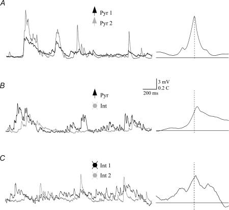
It is well established that sensory stimulation results in the activity of multiple functional columns in the neocortex. The manner in which neurones within each column are active in relation to each other is, however, not known. Multiple whole-cell recordings in activated neocortical slices from rat revealed diverse correlation profiles of excitatory synaptic input to different types of neurones. The specific correlation profile between any two neurones could be predicted by the settings of synaptic depression and facilitation at the input synapses. Simulations further showed that patterned activity is essential for synaptic dynamics to impose the temporal dispersion of excitatory input. We propose that synaptic dynamics choreograph neuronal activity within the neocortical microcircuit in a context-dependent manner.
Figures


Similar articles
-
Activity- and BDNF-induced plasticity of miniature synaptic currents in ES cell-derived neurons integrated in a neocortical network.J Neurophysiol. 2005 Dec;94(6):4538-43. doi: 10.1152/jn.00155.2005. J Neurophysiol. 2005. PMID: 16293594
-
Spontaneous and evoked synaptic rewiring in the neonatal neocortex.Proc Natl Acad Sci U S A. 2006 Aug 29;103(35):13214-9. doi: 10.1073/pnas.0604691103. Epub 2006 Aug 21. Proc Natl Acad Sci U S A. 2006. PMID: 16924105 Free PMC article.
-
Functional connectivity in layer IV local excitatory circuits of rat somatosensory cortex.J Neurophysiol. 2004 Oct;92(4):2137-50. doi: 10.1152/jn.01262.2003. Epub 2004 Jun 16. J Neurophysiol. 2004. PMID: 15201316
-
Does inhibition balance excitation in neocortex?Prog Biophys Mol Biol. 2005 Jan;87(1):109-43. doi: 10.1016/j.pbiomolbio.2004.06.008. Prog Biophys Mol Biol. 2005. PMID: 15471593 Review.
-
The neuronal transfer function: contributions from voltage- and time-dependent mechanisms.Prog Brain Res. 2007;165:1-12. doi: 10.1016/S0079-6123(06)65001-2. Prog Brain Res. 2007. PMID: 17925236 Review.
Cited by
-
Molecular and cellular approaches for diversifying and extending optogenetics.Cell. 2010 Apr 2;141(1):154-165. doi: 10.1016/j.cell.2010.02.037. Epub 2010 Mar 18. Cell. 2010. PMID: 20303157 Free PMC article.
-
Nicotinic ACh receptors in the hippocampus: role in excitability and plasticity.Nicotine Tob Res. 2012 Nov;14(11):1249-57. doi: 10.1093/ntr/nts091. Epub 2012 Apr 3. Nicotine Tob Res. 2012. PMID: 22472168 Free PMC article. Review.
-
Review of EEG Affective Recognition with a Neuroscience Perspective.Brain Sci. 2024 Apr 8;14(4):364. doi: 10.3390/brainsci14040364. Brain Sci. 2024. PMID: 38672015 Free PMC article. Review.
-
Basal forebrain innervation of the amygdala: an anatomical and computational exploration.Brain Struct Funct. 2025 Jan 13;230(1):30. doi: 10.1007/s00429-024-02886-1. Brain Struct Funct. 2025. PMID: 39805973 Free PMC article.
-
Synaptic input correlations leading to membrane potential decorrelation of spontaneous activity in cortex.J Neurosci. 2013 Sep 18;33(38):15075-85. doi: 10.1523/JNEUROSCI.0347-13.2013. J Neurosci. 2013. PMID: 24048838 Free PMC article.
References
-
- Anderson JS, Carandini M, Ferster D. Orientation tuning of input conductance, excitation, and inhibition in cat primary visual cortex. J Neurophysiol. 2000;84:909–926. - PubMed
-
- Beierlein M, Gibson JR, Connors BW. Two dynamically distinct inhibitory networks in layer 4 of the neocortex. J Neurophysiol. 2003;90:2987–3000. - PubMed
MeSH terms
LinkOut - more resources
Full Text Sources
Other Literature Sources
Miscellaneous