. 2004 Apr 15;556(Pt 2):415-28.
doi: 10.1113/jphysiol.2004.061762. Epub 2004 Feb 20.

Basolateral anion transport mechanisms underlying fluid secretion by mouse, rat and guinea-pig pancreatic ducts

Affiliations

Basolateral anion transport mechanisms underlying fluid secretion by mouse, rat and guinea-pig pancreatic ducts

M Paz Fernández-Salazar et al. J Physiol. .

Abstract

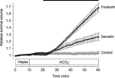
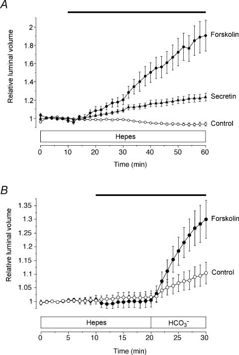
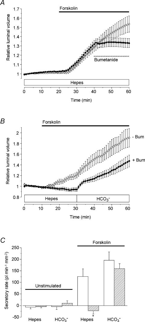
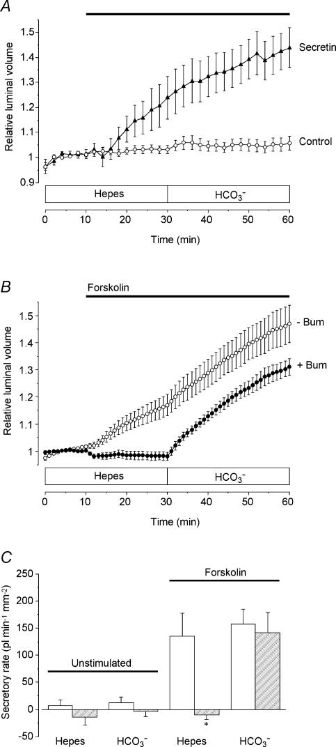
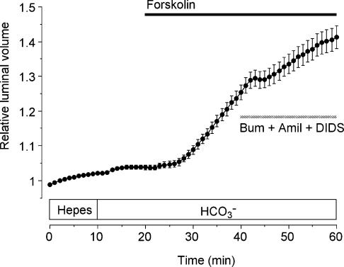
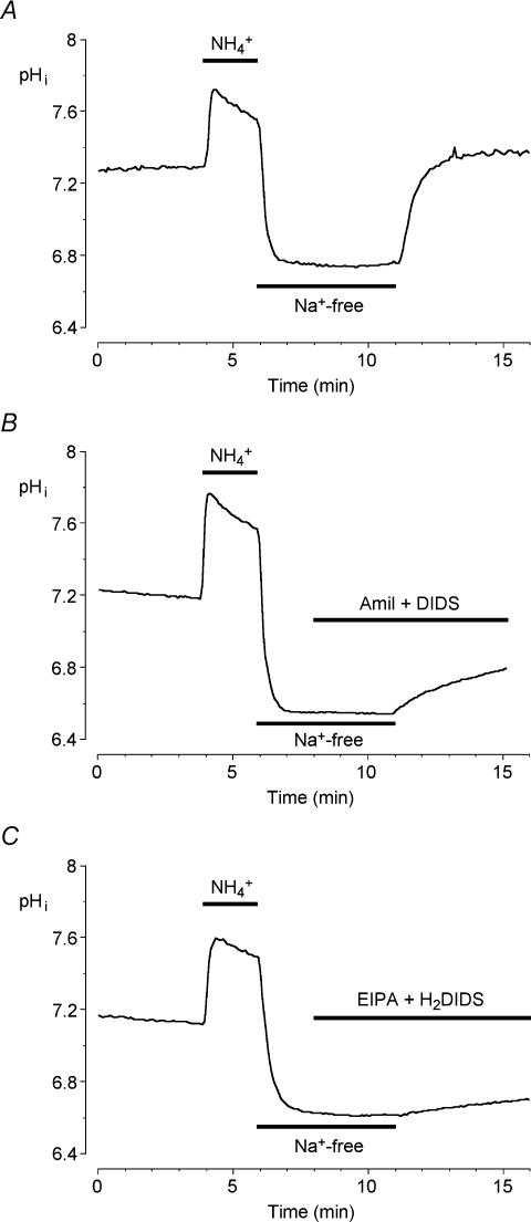
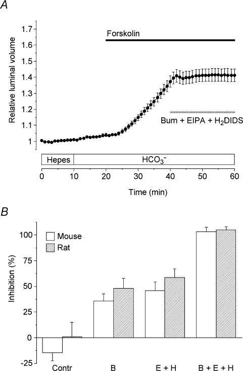
Fluid secretion by interlobular pancreatic ducts was determined by using video microscopy to measure the rate of swelling of isolated duct segments that had sealed following overnight culture. The aim was to compare the HCO(3)(-) requirement for secretin-evoked secretion in mouse, rat and guinea-pig pancreas. In mouse and rat ducts, fluid secretion could be evoked by 10 nm secretin and 5 microm forskolin in the absence of extracellular HCO(3)(-). In guinea-pig ducts, however, fluid secretion was totally dependent on HCO(3)(-). Forskolin-stimulated fluid secretion by mouse and rat ducts in the absence of HCO(3)(-) was dependent on extracellular Cl(-) and was completely inhibited by bumetanide (30 microm). It was therefore probably mediated by a basolateral Na(+)-K(+)-2Cl(-) cotransporter. In the presence of HCO(3)(-), forskolin-stimulated fluid secretion was reduced approximately 40% by bumetanide, approximately 50% by inhibitors of basolateral HCO(3)(-) uptake (3 microm EIPA and 500 microm H(2)DIDS), and was totally abolished by simultaneous application of all three inhibitors. We conclude that the driving force for secretin-evoked fluid secretion by mouse and rat ducts is provided by parallel basolateral mechanisms: Na(+)-H(+) exchange and Na(+)-HCO(3)(-) cotransport mediating HCO(3)(-) uptake, and Na(+)-K(+)-2Cl(-) cotransport mediating Cl(-) uptake. The absence or inactivity of the Cl(-) uptake pathway in the guinea-pig pancreatic ducts may help to account for the much higher concentrations of HCO(3)(-) secreted in this species.


Figures

Figure 1
Figure 1. Changes in relative luminal volume of mouse pancreatic ducts stimulated with secretin and forskolin
Mouse ducts superfused first with the Hepes-buffered solution were switched at 10 min to the HCO3-buffered solution for the remainder of the experiment. From 20 min the ducts were exposed either to no agonist (○, n = 64 in 9 experiments), to 10 nm secretin (▴, n = 10 in 5 experiments) or to 5 μm forskolin (•, n = 26 in 10 experiments) as indicated by the horizontal bar. Data are means ±s.e.m. of n values. The secretory rates given in the text were calculated from the rates of increase in relative luminal volume.
Figure 2
Figure 2. Fluid secretion by mouse and guinea-pig pancreatic ducts in the nominal absence of extracellular HCO3
A, mouse ducts superfused throughout with the Hepes-buffered solution and exposed either to no agonist (○, n = 4 in 3 experiments), to 10 nm secretin (▴, n = 9 in 6 experiments) or to 5 μm forskolin (•, n = 12 in 8 experiments) as indicated by the horizontal bar. B, guinea-pig ducts superfused first with the Hepes-buffered solution and then with the HCO3-buffered solution, and exposed either to no agonist (○, n = 22 in 8 experiments) or to 5 μm forskolin as indicated by the horizontal bar (•, n = 10 in 4 experiments).
Figure 3
Figure 3. Effect of bumetanide on fluid secretion by mouse pancreatic ducts in the presence or absence of HCO3
A, mouse ducts superfused throughout with the Hepes-buffered solution. From 20 min the ducts were stimulated with 5 μm forskolin (filled horizontal bar) and from 40 to 60 min they were exposed either to no blocker (○, n = 15 in 4 experiments) or to 30 μm bumetanide (•, n = 20 in 7 experiments) as indicated by the hatched horizontal bar. B, mouse ducts switched from the Hepes- to the HCO3-buffered solution at 30 min. From 10 min the ducts were stimulated with 5 μm forskolin (horizontal bar). Ducts were superfused either in the absence (○, n = 17 in 8 experiments) or in the continuous presence of 30 μm bumetanide (bum) (•, n = 11 in 6 experiments). C, mean secretory rates calculated from the data shown in Fig. 3B in ducts stimulated with 5 μm forskolin compared with equivalent control data obtained in unstimulated ducts (not shown; n = 12 in 4 experiments in the absence of bumetanide; n = 15 in 3 experiments in the presence of bumetanide). Secretory rates in the Hepes- and HCO3-buffered solutions were estimated over the 20–30 min and 50–60 min periods, respectively. Open bars represent values obtained in the absence of bumetanide, and hatched bars represent values obtained in ducts continuously superfused with 30 μm bumetanide. Secretory rates that in the presence of bumetanide were judged significantly different from the corresponding control values by unpaired Student's t test are indicated by an asterisk.
Figure 4
Figure 4. HCO3 dependence and bumetanide sensitivity of fluid secretion by rat pancreatic ducts
A, rat ducts superfused initially with the Hepes-buffered solution and after 30 min with the HCO3-buffered solution. From 10 min the ducts were exposed either to no agonist (○, n = 17 in 4 experiments) or to 10 nm secretin (▴, n = 10 in 4 experiments) as indicated by the horizontal bar. B, rat ducts superfused initially with the Hepes-buffered solution and after 30 min with the HCO3-buffered solution. From 10 min the ducts were exposed to 5 μm forskolin (horizontal bar), either in the absence (○, n = 14 in 4 experiments) or in the continuous presence of 30 μm bumetanide (bum) (•, n = 15 in 4 experiments). C, mean secretory rates calculated from the data shown in Fig. 4B in ducts stimulated with 5 μm forskolin compared with equivalent control data obtained in unstimulated ducts (not shown; n = 15 in 3 and 17 in 4 in the presence and absence of bumetanide, respectively). Secretory rates in the Hepes- and HCO3-buffered solutions were estimated over the 20–30 min and 50–60 min periods, respectively. Open bars represent values obtained in the absence of bumetanide, and hatched bars represent values obtained in ducts continuously superfused with 30 μm bumetanide. Secretory rates that in the presence of bumetanide were judged significantly different from the corresponding control values by unpaired Student's t test are indicated by an asterisk.
Figure 5
Figure 5. Effects of bumetanide, amiloride and DIDS on fluid secretion by mouse pancreatic ducts
Mouse ducts were stimulated with 5 μm forskolin in the presence of HCO3, as indicated by the filled horizontal bar, and then exposed to 30 μm bumetanide (bum), 300 μm amiloride (amil) and 100 μm DIDS as indicated by the hatched horizontal bar (n = 26 in 10 experiments).
Figure 6
Figure 6. Effects of transport inhibitors on intracellular pH recovery after an acid load in mouse pancreatic ductal cells
Mouse duct cells superfused with the HCO3-buffered solution were acid loaded by a brief (2 min) exposure to 20 mm NH4Cl followed by superfusion with a Na+-free solution for the following 5 min after which Na+ was restored. A, a representative control experiment performed in the absence of any inhibitors. B, pHi recovery in the presence of 300 μm amiloride (amil) and 100 μm DIDS, which were applied 3 min before Na+ was restored. C, pHi recovery in the presence of 3 μm EIPA and 500 μm H2DIDS. Traces are each representative of 5 different experiments.
Figure 7
Figure 7. Effects of bumetanide, EIPA and H2DIDS on fluid secretion by mouse and rat pancreatic ducts
A, mouse ducts were stimulated with 5 μm forskolin in the presence of HCO3 (filled horizontal bar) and then exposed to 30 μm bumetanide (bum), 3 μm EIPA and 500 μm H2DIDS as indicated by the hatched horizontal bar (n = 40 in 14 experiments). B, percentage inhibition of the secretory rate in mouse ducts (open bars) and rat ducts (hatched bars) in the absence of inhibitors (Contr; mouse n = 26 in 10, rat n = 14 in 7), in the presence of 30 μm bumetanide (B; mouse n = 35 in 11, rat n = 23 in 5), in the presence of 3 μm EIPA plus 500 μm H2DIDS (E + H; mouse n = 50 in 14, rat n = 45 in 9) and in the presence of all three inhibitors (B + E + H; mouse n = 40 in 14, rat n = 24 in 8). Values were calculated by comparing secretory rates in the 50–60 min period with those in the 30–40 min period in experiments which all followed the protocol shown in panel A.

Similar articles

Cited by

References

    1. Argent BE, Arkle S, Cullen MJ, Green R. Morphological, biochemical and secretory studies on rat pancreatic ducts maintained in tissue culture. Q J Exp Physiol. 1986;71:633–648. - PubMed
    1. Ashton N, Argent BE, Green R. Effect of vasoactive intestinal peptide, bombesin and substance P on fluid secretion by isolated rat pancreatic ducts. J Physiol. 1990;427:471–482. - PMC - PubMed
    1. Ashton N, Argent BE, Green R. Characteristics of fluid secretion from isolated rat pancreatic ducts stimulated with secretin and bombesin. J Physiol. 1991;435:533–546. - PMC - PubMed
    1. Ashton N, Evans RL, Elliott AC, Green R, Argent BE. Regulation of fluid secretion and intracellular messengers in isolated rat pancreatic ducts by acetylcholine. J Physiol. 1993;471:549–562. - PMC - PubMed
    1. Burghardt B, Elkjær ML, Kwon TH, Rácz GZ, Varga G, Steward MC, Nielsen S. Distribution of aquaporin water channels AQP1 and AQP5 in the ductal system of human pancreas. Gut. 2003;52:1008–1016. - PMC - PubMed

Publication types

MeSH terms

LinkOut - more resources