. 2004 May;15(5):2205-17.
doi: 10.1091/mbc.e03-07-0493. Epub 2004 Feb 20.

Regulation of dendritic branching and filopodia formation in hippocampal neurons by specific acylated protein motifs

Affiliations

Regulation of dendritic branching and filopodia formation in hippocampal neurons by specific acylated protein motifs

Catherine Gauthier-Campbell et al. Mol Biol Cell. 2004 May.

Abstract

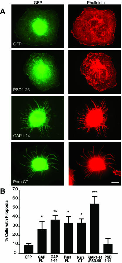
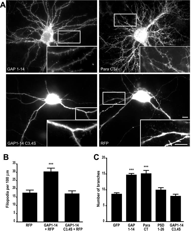
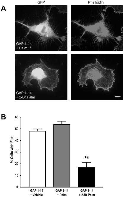
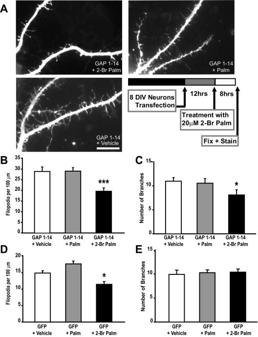
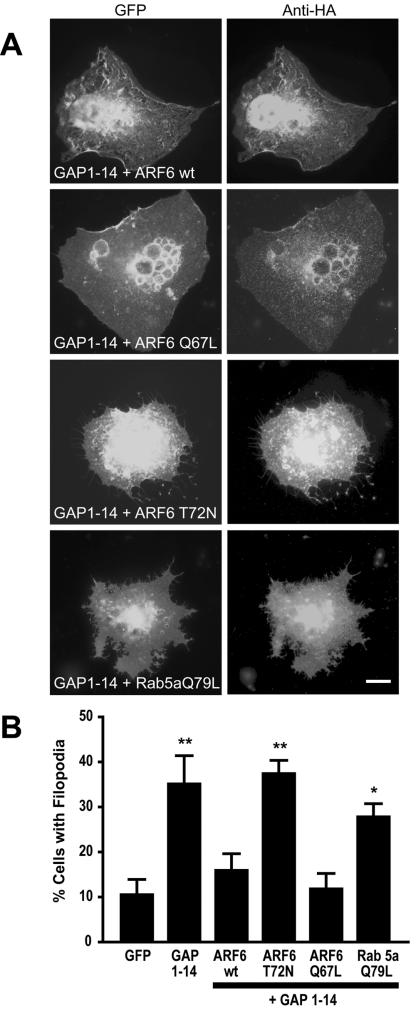
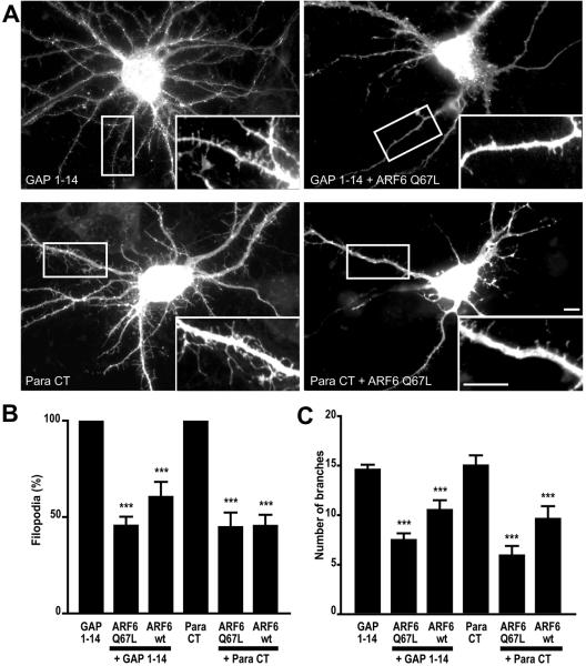
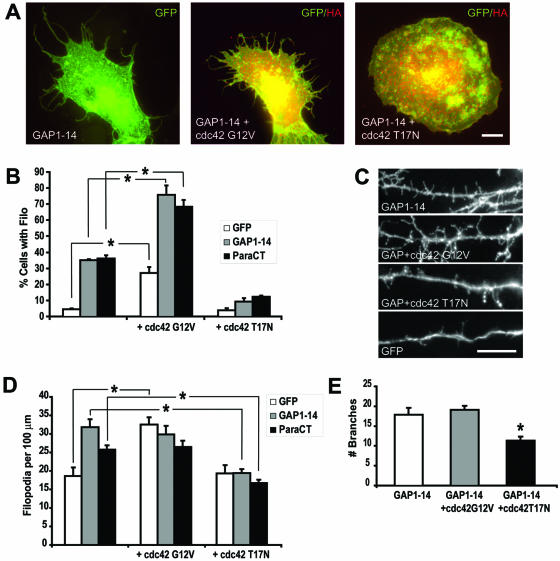
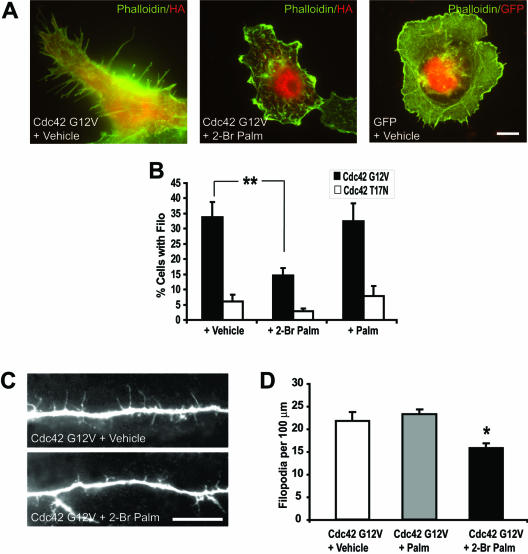
Although neuronal axons and dendrites with their associated filopodia and spines exhibit a profound cell polarity, the mechanism by which they develop is largely unknown. Here, we demonstrate that specific palmitoylated protein motifs, characterized by two adjacent cysteines and nearby basic residues, are sufficient to induce filopodial extensions in heterologous cells and to increase the number of filopodia and the branching of dendrites and axons in neurons. Such motifs are present at the N-terminus of GAP-43 and the C-terminus of paralemmin, two neuronal proteins implicated in cytoskeletal organization and filopodial outgrowth. Filopodia induction is blocked by mutations of the palmitoylated sites or by treatment with 2-bromopalmitate, an agent that inhibits protein palmitoylation. Moreover, overexpression of a constitutively active form of ARF6, a GTPase that regulates membrane cycling and dendritic branching reversed the effects of the acylated protein motifs. Filopodia induction by the specific palmitoylated motifs was also reduced upon overexpression of a dominant negative form of the GTPase cdc42. These results demonstrate that select dually lipidated protein motifs trigger changes in the development and growth of neuronal processes.


Figures

Figure 1.
Figure 1.
Filopodia induction in COS-7 cells. (A) COS-7 cells were transiently transfected with various constructs fused to GFP (green) as described in Table 1 and immunolabeled for F-actin with a rhodamine-conjugated phalloidin antibody (red). COS-7 cells transfected with full-length GAP-43 (GAP FL) and full-length paralemmin (Para FL) show increased branching vs. control GFP-transfected cells. When transfected with palmitoylation motifs of GAP-43 (GAP1–14) or of paralemmin (Para CT), COS-7 cells show extensive filopodial outgrowth, compared with cells transfected with the palmitoyaltion motif of PSD-95-GFP (PSD1–26). Filopodia induction was quantified by counting the number of cells showing extensive filopodia outgrowth (at least 5 filopodia of ≥10 μm per cell, or at least 20 filopodia of ≥5 μm) and expressed as a percent of cells “with filopodia.” (B) A graph showing that filopodial induction by GAP1–14, Para FL, Para CT, and GAP1–14/PSD-95 are statistically different from GFP (*p < 0.05; **p < 0.01; ***p < 0.01). Scale bar, 10 μm.
Figure 2.
Figure 2.
Filopodia induction and branching in neuronal cells. (A) Cultured hippocampal neurons transfected at day in vitro 7 (DIV 7) with either the palmitoylation motif of GAP-43 (GAP 1–14), or paralemmin (Para CT) show extensive branching and high filopodial density at DIV 10. In contrast, neurons transfected with RFP alone or RFP together with either the isolated palmitoylation motif of PSD-95 fused to GFP (PSD 1–26) or the palmitoylation-deficient motif of GAP-43 fused to GFP (GAP C3,4S) show less branching and lower filopodial density. Higher magnifications of dendritic branches and filopodia are shown in insets. (B) Graph shows the average number of filopodia per 100 μm and graph. (C) Graph shows the extent of branching in tranfected neurons with various constructs (***p < 0.001). Scale bars, 10 μm.
Figure 3.
Figure 3.
On-going palmitoylation of FIMs regulates filopodia formation in COS-7 cells. (A) COS-7 cells transfected with the GAP-43 palmitoylation motif and treated with 50 μM 2-bromopalmitate (24 h posttransfection), which blocks protein palmitoylation, show a significant decrease in filopodia outgrowth. (B) Graph shows the percentage of cells expressing filopodia for the different treatments; untreated and palmitate-treated cells are statistically different from 2-bromopalmitate-treated cells (**p < 0.01; ***p < 0.001). Scale bar, 10 μm.
Figure 4.
Figure 4.
Filopodia induction is palmitoylation-dependent and partly reversible in neuronal cells. Treating hippocampal neurons expressing the palmitoylation motif of GAP-43 (GAP 1–14) with 20 μM 2-bromopalmitate for 8 hs significantly reduces filopodia outgrowth. (A) Cells were transfected at DIV 8 and treatments with vehicle, palmitate (Palm) or 2-bromopalmitate (2-Br-Palm) were done 12 h posttransfection. (B) Graph shows the average number of filopodia per 100 μm for the different treatments; vehicle and palmitate-treated cells (GAP 1–14 + Palm) are statistically different from 2-bromopalmitate-treated cells (GAP 1–14 + 2-Br Palm; ***p < 0.001). (C) Graph shows the average number of branches for the different treatments; 2-bromopalmitate–treated cells are slightly but significantly different from vehicle and palmitate treated cells (*p < 0.05). (D) The total number of dendritic filopodia is reduced in GFP transfected neurons upon treatment with 2-Br-Palm treatment but not with Palm or Vehicle. (E) No significant change in dendritic branching was observed in GFP-transfected neurons upon treatment with 2-Br-Palm. Scale bar, 10 μm.
Figure 5.
Figure 5.
ARF6 regulates FIM-induced filopodia extension in COS-7 cells. (A) COS-7 cells cotransfected with the palmitoylation motif of GAP-43 and either wild-type ARF6 (ARF6wt), constitutively active form of ARF6 (ARF6-Q67L), dominant negative ARF6 (ARF6-T72N), or constitutively active Rab5a (Rab5a-Q79L). (B) Filopodia induction is quantified as the percentage of cells cotransfected showing filopodial outgrowth (*p < 0.05; **p < 0.01). Scale bar, 10 μm.
Figure 6.
Figure 6.
ARF6 regulation of dendritic branching and filopodia extension in neuronal cells. (A) Hippocampal neurons were cotransfected with the palmitoylation motif of GAP-43 (GAP 1–14) or the palmitoylation motif of paralemmin (Para CT) and either with wild-type ARF6 (ARF6wt) or with a constitutively active form of ARF6 (ARF6-Q67L). (B) Graph shows the relative filopodia density; data are expressed as a percentage of GAP1–14- or Para CT-transfected cells. (C) Graph shows the extent of branching in transfected neurons. (***p < 0.001). Scale bars, 10 μm.
Figure 7.
Figure 7.
Regulation of filopodia extension and dendritic branching by cdc42. (A) COS-7 cells were transfected with GFP, the palmitoylation motif of GAP-43 (GAP1–14) or the palmitoylation motif of paralemmin (ParaCT) and with either a constitutively active cdc42 (Cdc42 G12V) or a dominant negative cdc42 (Cdc42 T17N). Coexpression of GFP, GAP1–14, or ParaCT with Cdc42 G12V increased the number of cells with filopodia, whereas coexpression with Cdc42 T17N blocked filopodia induced by GAP1–14 and ParaCT. (B) Percentage of COS cells expressing filopodia for the different transfections (*p < 0.05). (C) Hippocampal neurons were cotransfected with GAP1–14 or GFP and either with Cdc42 G12V or Cdc42 T17N. (D) Graph shows the filopodia density for the various transfections. (E) Graph shows the extent of branching in transfected neurons. (*p < 0.05). Scale bars, 10 μm.
Figure 8.
Figure 8.
Palmitoylation modulates cdc42 induced filopodia extension and dendritic branching. (A) COS-7 cells were transfected with either a constitutively active or inactive cdc42 (Cdc42 G12V or Cdc42 T17N, respectively). Twelve hours posttransfection, cells were treated with either 20 μM palmitate (Palm) or 2-bromopalmitate (2-Br Palm) for 8 h. Results show a significant decrease in the number of cells with filopodia outgrowth compared with vehicle- or palmitate-treated cells. (B) Percentage of cells expressing filopodia for the different treatments; untreated and palmitate-treated cells are statistically different from 2-bromopalmitate–treated cells (**p < 0.01). (C) Images of hippocampal neurons illustrate changes in the density of dendritic filopodia in cells upon transfection with Cdc42 G12V and treatment with 20 μM 2-bromopalmitate or vehicle 12 h posttransfection. (D) Data show a significant decrease in filopodia density after treatment with 2-Br Palm. (*p < 0.05). Scale bars, 10 μm.

Similar articles

Cited by

References

    1. Aarts, L.H., Verkade, P., van Dalen, J.J., van Rozen, A.J., Gispen, W.H., Schrama, L.H., and Schotman, P. (1999). B-50/GAP-43 potentiates cytoskeletal reorganization in raft domains. Mol. Cell Neurosci. 14, 85–97. - PubMed
    1. Anderson, S.A., Classey, J.D., Conde, F., Lund, J.S., and Lewis, D.A. (1995). Synchronous development of pyramidal neuron dendritic spines and parvalbumin-immunoreactive chandelier neuron axon terminals in layer III of monkey prefrontal cortex. Neuroscience 67, 7–22. - PubMed
    1. Arni, S., Keilbaugh, S.A., Ostermeyer, A.G., and Brown, D.A. (1998). Association of GAP-43 with detergent-resistant membranes requires two palmitoylated cysteine residues. J. Biol. Chem. 273, 28478–28485. - PubMed
    1. Benowitz, L.I., and Routtenberg, A. (1997). GAP-43, an intrinsic determinant of neuronal development and plasticity. Trends Neurosci. 20, 84–91. - PubMed
    1. Bershadsky, A.D., and Futerman, A.H. (1994). Disruption of the Golgi apparatus by brefeldin A blocks cell polarization and inhibits directed cell migration. Proc. Natl. Acad. Sci. USA 91, 5686–5689. - PMC - PubMed

Publication types

MeSH terms