. 2004 May;15(5):2143-55.
doi: 10.1091/mbc.e03-09-0711. Epub 2004 Feb 20.

A role for sorting nexin 2 in epidermal growth factor receptor down-regulation: evidence for distinct functions of sorting nexin 1 and 2 in protein trafficking

Affiliations

A role for sorting nexin 2 in epidermal growth factor receptor down-regulation: evidence for distinct functions of sorting nexin 1 and 2 in protein trafficking

Anuradha Gullapalli et al. Mol Biol Cell. 2004 May.

Abstract

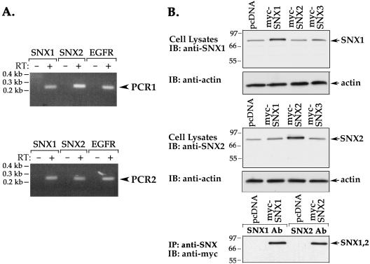
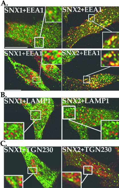
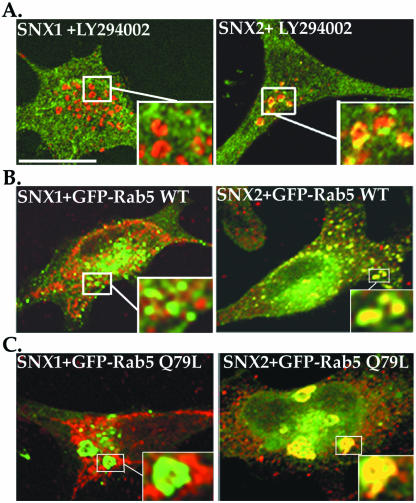
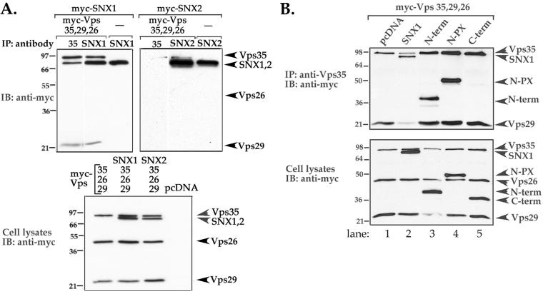
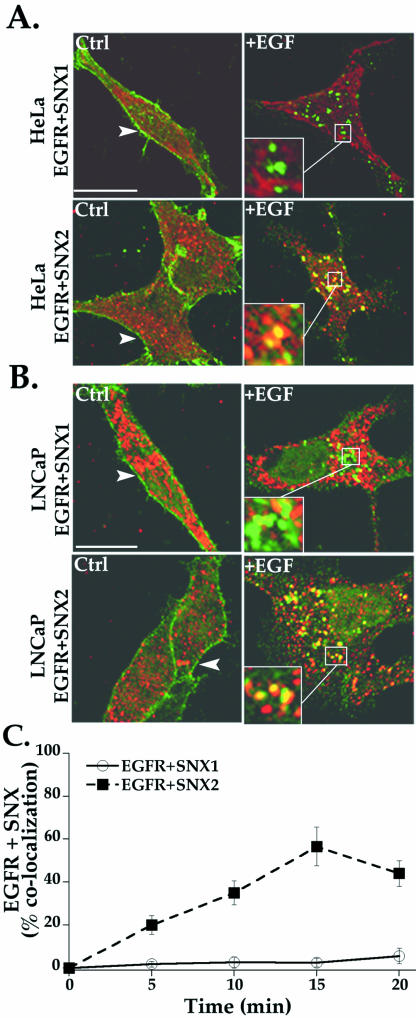
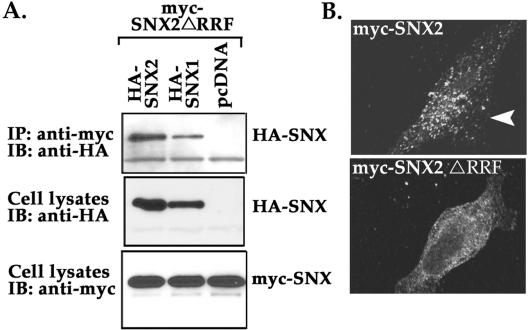
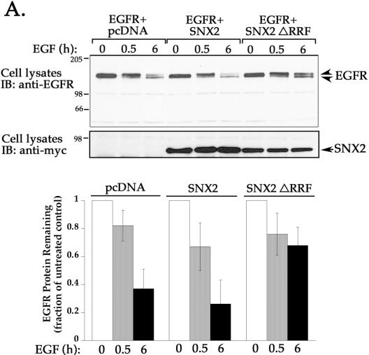
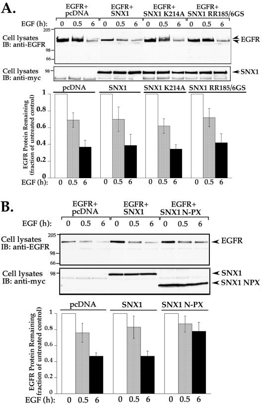
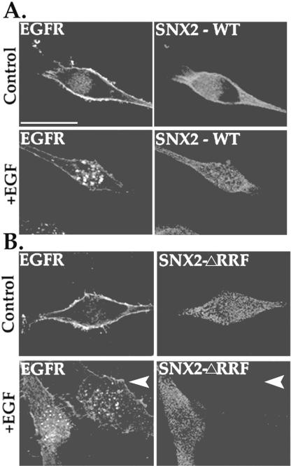
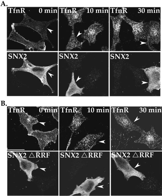
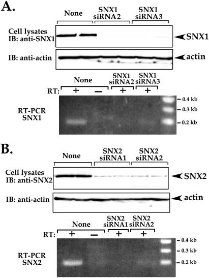
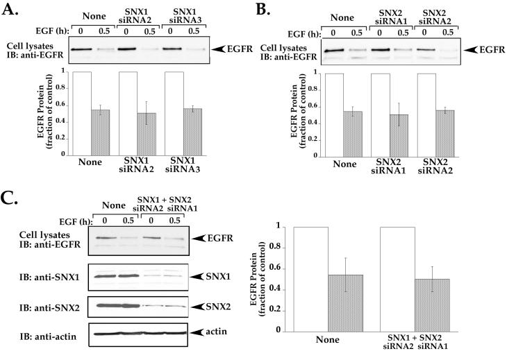
Sorting nexin 1 (SNX1) and SNX2, homologues of the yeast vacuolar protein-sorting (Vps)5p, contain a phospholipid-binding motif termed the phox homology (PX) domain and a carboxyl terminal coiled-coil region. A role for SNX1 in trafficking of cell surface receptors from endosomes to lysosomes has been proposed; however, the function of SNX2 remains unknown. Toward understanding the function of SNX2, we first examined the distribution of endogenous protein in HeLa cells. We show that SNX2 resides primarily in early endosomes, whereas SNX1 is found partially in early endosomes and in tubulovesicular-like structures distributed throughout the cytoplasm. We also demonstrate that SNX1 interacts with the mammalian retromer complex through its amino terminal domain, whereas SNX2 does not. Moreover, activated endogenous epidermal growth factor receptor (EGFR) colocalizes markedly with SNX2-positive endosomes, but minimally with SNX1-containing vesicles. To assess SNX2 function, we examined the effect of a PX domain-mutated SNX2 that is defective in vesicle localization on EGFR trafficking. Mutant SNX2 markedly inhibited agonist-induced EGFR degradation, whereas internalization remained intact. In contrast, SNX1 PX domain mutants failed to effect EGFR degradation, whereas a SNX1 deletion mutant significantly inhibited receptor down-regulation. Interestingly, knockdown of SNX1 and SNX2 expression by RNA interference failed to alter agonist-induced EGFR down-regulation. Together, these findings suggest that both SNX1 and SNX2 are involved in regulating lysosomal sorting of internalized EGFR, but neither protein is essential for this process. These studies are the first to demonstrate a function for SNX2 in protein trafficking.


Figures

Figure 1.
Figure 1.
SNX1 and SNX2 are expressed endogenously in HeLa cells. (A) Expression of SNX1, SNX2, and EGFR mRNAs in HeLa cells was detected by RT-PCR. Total cellular RNA (2 μg) was reverse-transcribed and PCR amplified. Mock RT-PCR of total cellular RNA with no reverse transcriptase added is shown in lanes marked as RT: –. SNX1 and SNX2 mRNA was analyzed twice by RT-PCR by using two distinct sets of primers and revealed the presence of mRNA transcript in HeLa cells. (B) HeLa cells transiently transfected with either myc-tagged SNX1, SNX2, SNX3, or pcDNA vector were lysed and immunoblotted with rabbit polyclonal anti-SNX1 or anti-SNX2 antibodies (top and middle, respectively). Actin expression was also detected as a loading control. HeLa cells transiently expressing either myc-SNX1, SNX2, or pcDNA vector were lysed and immunoprecipitated with anti-SNX polyclonal antibodies, respectively. Immunoprecipitates were immunoblotted for SNXs by using chicken anti-myc antibody (bottom).
Figure 2.
Figure 2.
Endogenous SNX1 and SNX2 localize largely to distinct intracellular compartments. (A–C) HeLa cells were fixed, permeabilized, and immunostained for endogenous SNX1 or SNX2 (green) and either EEA1, LAMP1, or TGN230 (shown in red) and examined by confocal microscopy. These images are representative of many cells examined in at least five independent experiments. Note the prominent colocalization (yellow) of endogenous SNX2 and EEA1 shown in the merged image (A), and the partial colocalization between SNX1 and EEA1. Neither endogenous SNX1 nor SNX2 showed significant colocalization with LAMP1 or TGN230 (B and C). The insets are magnifications of boxed areas. Bar, 10 μm.
Figure 3.
Figure 3.
Endogenous SNX2 localization is altered by inhibition of PI-3 kinase activity and expression of Rab5 Q79L mutant. (A) HeLa cells were incubated in the absence (our unpublished data) or presence of 50 μM LY294002 compound for 1 h at 37°C, fixed, permeabilized, and immunostained for endogenous SNX1 or SNX2 (green) and EEA1 (red). Colocalization was assessed by confocal microscopy. Note the localization of endogenous SNX2 in enlarged endosomes observed in LY294002 PI-3 kinase inhibitor treated cells. (B and C) HeLa cells were transiently transfected with GFP-tagged wild-type (WT) or mutant Q79L Rab5, fixed, permeabilized, and immunostained for endogenous SNX1 or SNX2 (red). Insets are magnifications of boxed areas. Note the prominent colocalization (yellow) of endogenous SNX2 with both wild-type and Q79L mutant Rab5 as detected by confocal microscopy. These images are representative of many cells observed in three independent experiments. Bar, 10 μm.
Figure 4.
Figure 4.
SNX1 amino-terminal domain mediates interaction with the retromer complex in mammalian cells. (A) HeLa cells transiently expressing the retromer subunits (myc-tagged Vps35, Vps29, and Vps26) together with either myc-tagged SNX1 or SNX2 were lysed and immunoprecipitated with either anti-Vps35, -SNX1, or -SNX2 rabbit polyclonal antibodies. Immunoprecipitates were resolved by SDS-PAGE and then immunoblotted with chicken anti-myc antibody (top panels). The expression of SNXs and Vps subunits in total cell lysates was detected with anti-myc antibody (bottom). Note the robust association between SNX1 and Vps35 and the lack of association with SNX2 (top right). Similar findings were observed in three separate experiments. (B) HeLa cells transiently transfected with myc-tagged Vps35, Vps29, Vps26, and either full-length SNX1, deletion mutants of SNX1, or pcDNA vector alone were lysed, immunoprecipitated with anti-Vps35 antibody, resolved by SDS-PAGE, and immunoblotted with anti-myc antibody to detect protein expression (top). The expression of SNX1, deletion mutants, and Vps subunits in total cell lysates was detected with anti-myc antibody (bottom). Note that the amino terminus alone is sufficient to interact with the retromer complex (lane 3).
Figure 5.
Figure 5.
Colocalization of activated endogenous EGFR with SNX2. (A) Serum-starved HeLa and (B) LNCaP cells were incubated in the absence or presence of 100 ng/ml EGF for 20 min at 37°C. Cells were fixed, permeabilized, and immunostained for endogenous EGFR (green) and SNXs (red) and examined by confocal microscopy. The colocalization of activated EGFR with SNX2 was easily detected in HeLa and LNCaP cells, whereas EGFR and SNX1 showed minimal overlap in either cell type. Similar findings were observed in three separate experiments. Insets are magnifications of boxed areas. Bar, 10 μm. (C) Results of quantitative analysis are expressed as a percentage of HeLa cells that showed EGFR-positive vesicles and costained for either SNX1 or SNX2 after various times of agonist treatment. The data (mean ± SEM) are representative of three separate experiments.
Figure 6.
Figure 6.
SNX2 mutant can self-associate but fails to localize to an endosomal compartment. (A) HeLa cells transiently cotransfected with myc-SNX2 ΔRRF mutant together with wild-type HA-SNX2, HA-SNX1, or pcDNA vector were lysed and immunoprecipitated with anti-myc antibody. Immunoprecipitates were immunoblotted for HA-SNXs by using anti-HA peroxidase conjugated antibody (top). The expression of HA-SNXs and myc-SNX2 mutant in total cell lysates is shown in the middle and bottom panels, respectively. (B) HeLa cells transiently overexpressing wild-type and SNX2 mutant protein were permeabilized with saponin and endosomal localization detected by immunofluorescence microscopy. Note the loss of endosomal localization of SNX2 ΔRRF mutant compared with wild-type SNX2.
Figure 7.
Figure 7.
Overexpression of SNX2 mutant markedly inhibits agonist-induced EGFR degradation. HeLa cells transiently cotransfected with EGFR and either pcDNA vector, SNX2, or SNX2 ΔRRF mutant were serum-starved and incubated in the absence or presence of EGF ligand (100 ng/ml) for 0, 0.5, or 6 h at 37°C. Cells were then lysed, and equivalent amounts of lysates were resolved by SDS-PAGE. The amount of EGFR remaining was detected with anti-EGFR antibody (top) and quantitated by Fluor-S Imager. The expression of SNX2 wild type and ΔRRF mutant in cell lysates was detected with anti-myc antibody (bottom). Results in the bar graph are expressed as a fraction of EGFR measured in untreated control lysates and was determined for each transfection condition. The data are represented as the mean ± SEM of three separate experiments. Note the marked inhibition of agonist-induced EGFR degradation in cells expressing SNX2 ΔRRF mutant.
Figure 8.
Figure 8.
Effect of SNX1 PX domain and deletion mutants on agonist-stimulated EGFR degradation. (A and B) Serum-deprived HeLa cells transiently cotransfected with EGFR and either wild-type or mutant SNX1, or vector only were treated and processed as described in Figure 7. The expression of wild type and SNX1 mutants in cell lysates was detected with anti-myc antibody. The data are shown as the mean ± SEM of at least three independent experiments. Note that SNX1 PX domain mutants fail to inhibit agonist-induced EGFR degradation, whereas SNX1 N-PX deletion mutant (1–273) blocked receptor degradation.
Figure 9.
Figure 9.
Agonist-induced EGFR internalization in SNX2 wild-type and mutant expressing cells. HeLa cells transiently cotransfected with GFP-tagged EGFR and either (A) SNX2 wild-type (WT) or (B) ΔRRF mutant were serum deprived and then treated in the absence (control) or presence of 100 ng/ml EGF for 20 min at 37°C. Cells were fixed, immunostained for SNX2 expression, and imaged by confocal microscopy. Note the presence of EGFR-positive endosomes in both SNX2 wild-type and ΔRRF mutant-transfected cells. These results are representative of many cells examined in at least three individual experiments. Bar, 10 μm.
Figure 10.
Figure 10.
Effect of wild-type and mutant SNX2 on TfnR trafficking. (A and B) HeLa cells transiently transfected with either SNX2 wild type or mutant were serum starved and incubated with anti-TfnR antibody for 1 h at 4°C. Cells were washed to remove unbound antibody and then incubated in medium for 0, 10, or 30 min at 37°C. Cells were then fixed, permeabilized, and immunostained for TfnR and SNX2 and processed for microscopy. The arrowheads indicate cells transfected with either wild-type or mutant SNX2. The findings are representative of many cells examined in two independent experiments. Bar, 10 μm.
Figure 11.
Figure 11.
SNX1 and SNX2 knockdown by siRNA in HeLa cells. (A and B) HeLa cells were transiently transfected with 100 nM siRNA targeted to either SNX1 or SNX2 mRNA sequences or mock transfected indicated as “None.” Cell lysates were prepared and immunoblotted with either anti-SNX1 or anti-SNX2 antibodies (top panels). Actin expression was also detected as a loading control. Total cellular RNA was also prepared from siRNA transfected cells and reverse-transcribed and PCR amplified. A mock RT-PCR of total cellular RNA with no reverse transcriptase added is shown in lanes marked as RT: –.
Figure 12.
Figure 12.
Knockdown of SNX1 and SNX2 protein expression fails to affect agonist-induced EGFR degradation. (A and B) HeLa cells were transiently transfected with either 100 nM SNX1 or SNX2-specific siRNAs as described above and then incubated in the absence or presence of EGF (100 ng/ml) for 0.5 h at 37°C. “None” indicates cells that were mock transfected. Cell lysates were prepared and the amount of endogenous EGFR remaining was detected by immunoblot and quantitated as described above. The data shown (mean ± SEM) are representative of three independent experiments performed in duplicate. (C) HeLa cells were transiently transfected with 100 nM SNX1 and SNX2 siRNAs to achieve knockdown of both proteins. Cells were then incubated with 100 ng/ml EGF for 0.5 h at 37°C, lysates were prepared and immunblotted as described. The extent of EGFR degradation was quantitated and shown as fraction of control. Note that neither SNX1 nor SNX2 seems to be essential for agonist-induced EGFR down-regulation.

References

    1. Bishop, N., Horman, A., and Woodman, P. (2002). Mammalian class E vps proteins recognize ubiquitin and act in the removal of endosomal protein-ubiquitin conjugates. J. Cell Biol. 157, 91–101. - PMC - PubMed
    1. Bravo, J., et al. (2001). The crystal structure of the PX domain from p40phox bound to phosphatidylinositol 3-phosphate. Mol. Cell. 8, 829–839. - PubMed
    1. Brown, W.J., DeWald, D.B., Emr, S.D., Plutner, H., and Balch, W.E. (1995). Role for phosphatidylinositol 3-kinase in the sorting and transport of newly synthesized lysosomal enzymes in mammalian cells. J. Cell Biol. 130, 781–796. - PMC - PubMed
    1. Carter, R.E., and Sorkin, A. (1998). Endocytosis of functional epidermal growth factor receptor-green fluorescent protein chimera. J. Biol. Chem. 273, 35000–35007. - PubMed
    1. Chin, L.S., Raynor, M.C., Wei, X., Chen, H.Q., and Li, L. (2001). Hrs interacts with sorting nexin 1 and regulates degradation of epidermal growth factor receptor. J. Biol. Chem. 276, 7069–7078. - PubMed

Publication types

MeSH terms