. 2004 Mar 2;101(9):2975-80.
doi: 10.1073/pnas.0400093101. Epub 2004 Feb 20.

Disruption of forkhead transcription factor (FOXO) family members in mice reveals their functional diversification

Affiliations

Disruption of forkhead transcription factor (FOXO) family members in mice reveals their functional diversification

Taisuke Hosaka et al. Proc Natl Acad Sci U S A. .

Abstract

Genetic analysis in Caenorhabditis elegans has uncovered essential roles for DAF-16 in longevity, metabolism, and reproduction. The mammalian orthologs of DAF-16, the closely-related FOXO subclass of forkhead transcription factors (FKHR/FOXO1, FKHRL1/FOXO3a, and AFX/FOXO4), also have important roles in cell cycle arrest, apoptosis and stress responses in vitro, but their in vivo physiological roles are largely unknown. To elucidate their role in normal development and physiology, we disrupted each of the Foxo genes in mice. Foxo1-null embryos died on embryonic day 10.5 as a consequence of incomplete vascular development. Foxo1-null embryonic and yolk sac vessels were not well developed at embryonic day 9.5, and Foxo1 expression was found in a variety of embryonic vessels, suggesting a crucial role of this transcription factor in vascular formation. On the other hand, both Foxo3a- and Foxo4-null mice were viable and grossly indistinguishable from their littermate controls, indicating dispensability of these two members of the Foxo transcription factor family for normal vascular development. Foxo3a-null females showed age-dependent infertility and had abnormal ovarian follicular development. In contrast, histological analyses of Foxo4-null mice did not identify any consistent abnormalities. These results demonstrate that the physiological roles of Foxo genes are functionally diverse in mammals.


Figures
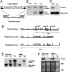
Fig. 4.
Fig. 4.
Targeted disruption of the Foxo3a gene and Foxo4 gene. (A) Targeting strategy for Foxo3a. A retroviral promoter trap vector was inserted in intron 1 of Foxo3a. SA, splice acceptor site; SD, splice donor site; LTR, long terminal repeat sequence; pA, polyadenylation signal; puro, puromycin. (B) Southern analysis of SacI-digested genomic DNA obtained from a mating of Foxo3a+/- mice by using the Neo- and Csk-specific probes. Different intensities demonstrated by hybridization of the Neo-specific probe discriminate zero, one, or two Foxo3a gene disruptions, representing +/+, +/-, and -/- mice, respectively. (C) Western analysis of lung tissue lysates from Foxo3a+/+ and Foxo3a-/- mice by using antibodies against Foxo3a and β-actin. (D) Restriction maps of the wild-type Foxo4 locus, the targeting vector, and the targeted locus. The targeting vector contained a PGK-Neo cassette instead of the Foxo4 exon1. A targeting probe (3′ region of Foxo4 cDNA) was also indicated. Restriction enzymes were indicated as B, BamHI; C, ClaI; E, EcoRI; H, HindIII; N, NcoI; and Sp, SpeI. (E) Southern analysis of BamHI-digested genomic DNA obtained from Foxo4 female (+/+, +/-, and -/-) and male (+/y and -/y) mice by using a probe from the 3′ region of Foxo4 cDNA. The Foxo4 wild-type allele (10 kb) and targeted allele (16 kb) were indicated. (F) Northern analysis of total RNA from skeletal muscle of Foxo4 female (+/+ and -/-) and male (+/y and -/y) mice by using the same probe used for Southern blot analysis. The ethidium bromide-staining 28S and 18S bands are also indicated.
Fig. 1.
Fig. 1.
Disruption of the Foxo1 gene results in embryonic lethality. (A) PCR analysis with genomic DNA from E9.5 yolk sacs obtained from a mating of Foxo1+/- mice. PCR amplification from the wild-type and Foxo1-targeted loci resulted in fragment sizes of 301 bp and 815 bp, respectively. Samples were independently amplified with each primer set. (B) Western blot analysis of lysates from E9.5 Foxo1+/+ and Foxo1-/- embryos by using antibodies against Foxo1 and β-actin. (C) At E9.5, both Foxo1+/+ and Foxo1+/- yolk sacs had well developed blood vessels, whereas Foxo1-/- yolk sacs lacked them. (D) At E9.5, Foxo1+/+ and Foxo1+/- embryos were phenotypically indistinguishable in appearance, whereas Foxo1-/- embryos were approximately half their size. In addition, cardiac looping was retarded and the pericardium was distended (arrowhead). (Scale bar, 500 μm.)
Fig. 2.
Fig. 2.
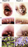
Foxo1-/- embryos and yolk sacs show defective vascular development. (A) PECAM-1 immunostaining of whole-mount E9.5 Foxo1+/+ and Foxo1-/- embryos, respectively. Note the thin and disorganized dorsal aorta in Foxo1-/- embryo compared with Foxo1+/+ embryo (arrowheads). (B) Magnified view of the E9.5 Foxo1+/+ and Foxo1-/- intersomitic vessels. Intersomitic vessels in Foxo1-/- embryo were disorganized compared with Foxo1+/+ (arrowheads). da, dorsal aorta. (C) Magnified view of the E9.5 Foxo1+/+ and Foxo1-/- head vessels. The vessels of the head, including the branches of the internal carotid artery, were properly developed in Foxo1+/+ but not in Foxo1-/- embryos (arrowheads). (D) PECAM-1 staining of Foxo1+/+ and Foxo1-/- yolk sacs. Properly developed vasculature was present in Foxo1+/+ yolk sacs but not in Foxo1-/- yolk sacs.
Fig. 3.
Fig. 3.
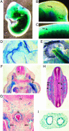
LacZ expression from the insertional targeting vector in heterozygous Foxo+/- embryos and extraembryonic tissues at E11.5. (A) Whole-mount lacZ staining of embryo. LacZ was expressed in several tissues such as the somites (so), branchial arch (ba), heart (h), endolymphatic diverticulum of otocyst (ed), and trigeminal ganglia (V). (B and C) Magnified view of the lacZ stained head and trunk, respectively. Lac Z staining was found in branches of the internal carotid artery (ica), anterior cardinal vein (acv), and intersomitic vessels (isv). (D and E) Whole-mount lacZ staining of the yolk sac and umbilical cord, respectively. Both vitelline vessels and both umbilical vessels were stained. (F-H) LacZ staining for sectioned E11.5 embryo. Various vessels including the capillaries in the neural tube (nt), dorsal aorta (da), posterior cardinal vein (pcv) and intersomitic vessels (isv) were stained. (G) Magnified view of F. so, somite; nc, notocord. (I) Section of lacZ-stained umbilical cord. The endothelial layers of the two vessels (common umbilical artery and vein) were stained.
Fig. 5.
Fig. 5.
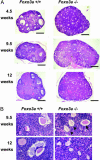
Histological analysis of ovaries with hematoxylin/eosin staining. (A) At 4.5 weeks of age, developing follicles containing growing oocytes but no antrums (type 3b-5b) were prominent, but mature follicles containing antrums (type 6-7) were less common in Foxo3a-/- ovaries. At 9.5 weeks of age, Foxo3a-/- ovaries had many developing follicles and few mature follicles, similar to those at 4.5 weeks. At 12 weeks of age, Foxo3a-/- ovaries had no developing follicles, and all oocytes had undergone degeneration. Each specimen was sectioned at its largest diameter. (Scale bar, 500 μm.) (B) Various stages of follicles were found in wild-type ovaries at both 9.5 and 12 weeks of age. Normal oocytes (arrow) were present, but degenerating oocytes (arrowheads) were prominent in Foxo3a-/- ovaries at 9.5 weeks of age. All oocytes had undergone degeneration in Foxo3a-/- ovaries at 12 weeks of age.

Similar articles

Cited by

References

    1. Arden, K. C. & Biggs, W. H., III (2002) Arch. Biochem. Biophys. 403, 292-298. - PubMed
    1. Lin, K., Dorman, J. B., Rodan, A. & Kenyon, C. (1997) Science 278, 1319-1322. - PubMed
    1. Ogg, S., Paradis, S., Gottlieb, S., Patterson, G. I., Lee, L., Tissenbaum, H. A. & Ruvkun, G. (1997) Nature 389, 994-999. - PubMed
    1. Tissenbaum, H. A. & Ruvkun, G. (1998) Genetics 148, 703-717. - PMC - PubMed
    1. Biggs, W. H., III, Meisenhelder, J., Hunter, T., Cavenee, W. K. & Arden, K. C. (1999) Proc. Natl. Acad. Sci. USA 96, 7421-7426. - PMC - PubMed

Publication types