

Laser light-scattering evidence for an altered association of beta B1-crystallin deamidated in the connecting peptide
- PMID: 14978307
- PMCID: PMC2286738
- DOI: 10.1110/ps.03427504
Laser light-scattering evidence for an altered association of beta B1-crystallin deamidated in the connecting peptide
Abstract
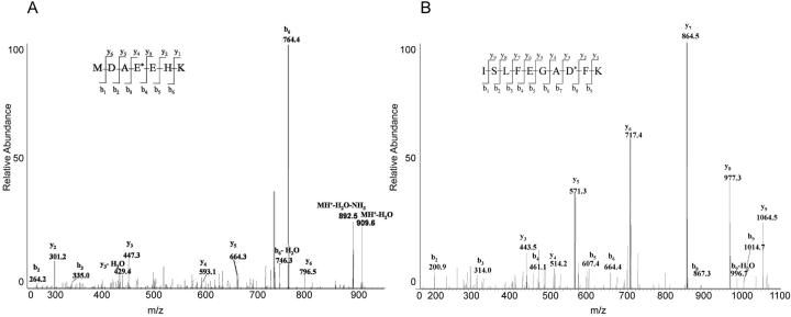
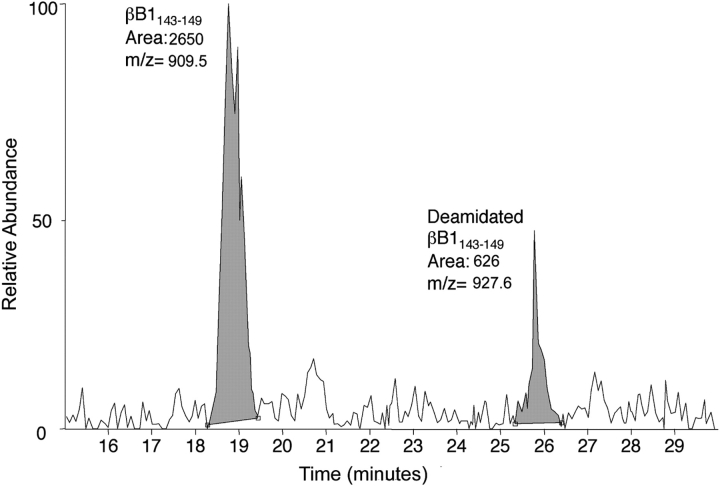
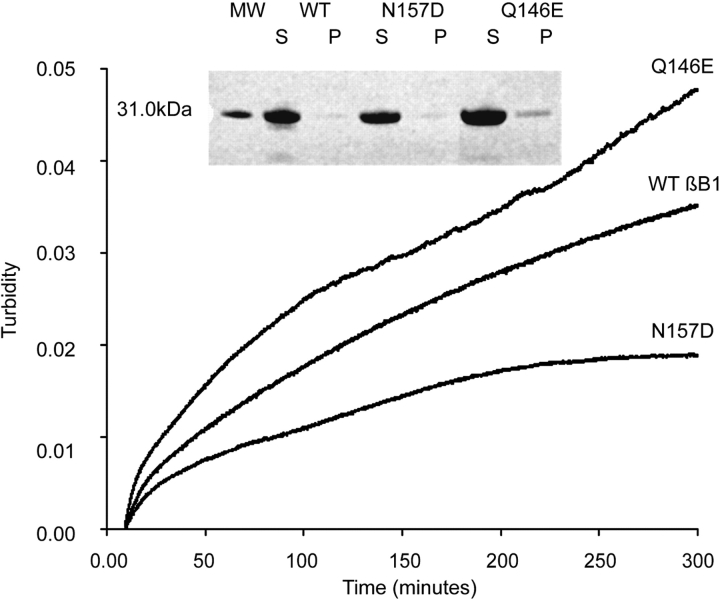
Deamidation is a prevalent modification of crystallin proteins in the vertebrate lens. The effect of specific sites of deamidation on crystallin stability in vivo is not known. Using mass spectrometry, a previously unreported deamidation in beta B1-crystallin was identified at Gln146. Another deamidation was investigated at Asn157. It was determined that whole soluble beta B1 contained 13%-17% deamidation at Gln146 and Asn157. Static and quasi-elastic laser light scattering, circular dichroism, and heat aggregation studies were used to explore the structure and associative properties of recombinantly expressed wild-type (wt) beta B1 and the deamidated beta B1 mutants, Q146E and N157D. Dimer formation occurred for wt beta B1, Q146E, and N157D in a concentration-dependent manner, but only Q146E showed formation of higher ordered oligomers at the concentrations studied. Deamidation at Gln146, but not Asn157, led to an increased tendency of beta B1 to aggregate upon heating. We conclude that deamidation creates unique effects depending upon where the deamidation is introduced in the crystallin structure.
Figures


Similar articles
-
Deamidation alters interactions of beta-crystallins in hetero-oligomers.Mol Vis. 2009;15:241-9. Epub 2009 Jan 28. Mol Vis. 2009. PMID: 19190732 Free PMC article.
-
Deamidation alters the structure and decreases the stability of human lens betaA3-crystallin.Biochemistry. 2007 Jul 31;46(30):8861-71. doi: 10.1021/bi700487q. Epub 2007 Jul 7. Biochemistry. 2007. PMID: 17616172 Free PMC article.
-
Quantitative measurement of deamidation in lens betaB2-crystallin and peptides by direct electrospray injection and fragmentation in a Fourier transform mass spectrometer.Mol Vis. 2005 Dec 28;11:1211-9. Mol Vis. 2005. PMID: 16402021 Free PMC article.
-
Lens β-crystallins: the role of deamidation and related modifications in aging and cataract.Prog Biophys Mol Biol. 2014 Jul;115(1):21-31. doi: 10.1016/j.pbiomolbio.2014.02.004. Epub 2014 Mar 6. Prog Biophys Mol Biol. 2014. PMID: 24613629 Free PMC article. Review.
-
Alpha crystallin: the quest for a homogeneous quaternary structure.Exp Eye Res. 2009 Feb;88(2):190-4. doi: 10.1016/j.exer.2008.07.007. Epub 2008 Jul 25. Exp Eye Res. 2009. PMID: 18703051 Free PMC article. Review.
Cited by
-
Age-related changes in human crystallins determined from comparative analysis of post-translational modifications in young and aged lens: does deamidation contribute to crystallin insolubility?J Proteome Res. 2006 Oct;5(10):2554-66. doi: 10.1021/pr050473a. J Proteome Res. 2006. PMID: 17022627 Free PMC article.
-
Identification of crystallin modifications in the human lens cortex and nucleus using laser capture microdissection and CyDye labeling.Mol Vis. 2010 Mar 23;16:476-94. Mol Vis. 2010. PMID: 20352024 Free PMC article.
-
Deamidation destabilizes and triggers aggregation of a lens protein, betaA3-crystallin.Protein Sci. 2008 Sep;17(9):1565-75. doi: 10.1110/ps.035410.108. Epub 2008 Jun 20. Protein Sci. 2008. PMID: 18567786 Free PMC article.
-
Structural and functional roles of deamidation of N146 and/or truncation of NH2- or COOH-termini in human αB-crystallin.Mol Vis. 2011;17:2407-20. Epub 2011 Sep 14. Mol Vis. 2011. PMID: 21976952 Free PMC article.
-
Ubiquitin proteasome pathway-mediated degradation of proteins: effects due to site-specific substrate deamidation.Invest Ophthalmol Vis Sci. 2010 Aug;51(8):4164-73. doi: 10.1167/iovs.09-4087. Epub 2010 Jun 30. Invest Ophthalmol Vis Sci. 2010. PMID: 20592226 Free PMC article.
References
-
- Bateman, O.A., Lubsen, N.H., and Slingsby, C. 2001. Association behaviour of human βB1-crystallin and its truncated forms. Exp. Eye Res. 73 321–331. - PubMed
-
- Bax, B., Lapatto, R., Nalini, V., Driessen, H., Lindley, P.F., Mahadevan, D., Blundell, T.L., and Slingsby, C. 1990. X-ray analysis of β B2-crystallin and evolution of oligomeric lens proteins. Nature 347 776–780. - PubMed
-
- Bindels, J.G., Koppers, A., and Hoenders, H.J. 1981. Structural aspects of bovine β-crystallins: Physical characterization including dissociation–association behavior. Exp. Eye Res. 33 333–343. - PubMed
-
- Blundell, T., Lindley, P., Miller, L., Moss, D., Slingsby, C., Tickle, I., Turnell, B., and Wistow, G. 1981. The molecular structure and stability of the eye lens: X-ray analysis of γ-crystallin II. Nature 289 771–777. - PubMed
-
- Cantor, C.R. and Schimmel, P.R. 1980. Biophysical chemistry. W.H. Freeman, San Francisco.
Publication types
MeSH terms
Substances
Grants and funding
LinkOut - more resources
Full Text Sources
