. 2004;6(2):R116-29.
doi: 10.1186/bcr757. Epub 2004 Jan 15.

Malignant myoepithelial cells are associated with the differentiated papillary structure and metastatic ability of a syngeneic murine mammary adenocarcinoma model

Affiliations

Malignant myoepithelial cells are associated with the differentiated papillary structure and metastatic ability of a syngeneic murine mammary adenocarcinoma model

Viviana Bumaschny et al. Breast Cancer Res. 2004.

Abstract

Background: The normal duct and lobular system of the mammary gland is lined with luminal and myoepithelial cell types. Although evidence suggests that myoepithelial cells might suppress tumor growth, invasion and angiogenesis, their role remains a major enigma in breast cancer biology and few models are currently available for exploring their influence. Several years ago a spontaneous transplantable mammary adenocarcinoma (M38) arose in our BALB/c colony; it contains a malignant myoepithelial cell component and is able to metastasize to draining lymph nodes and lung.

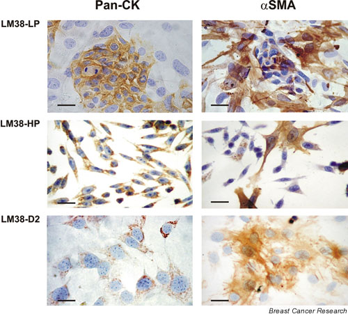
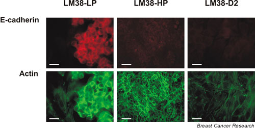
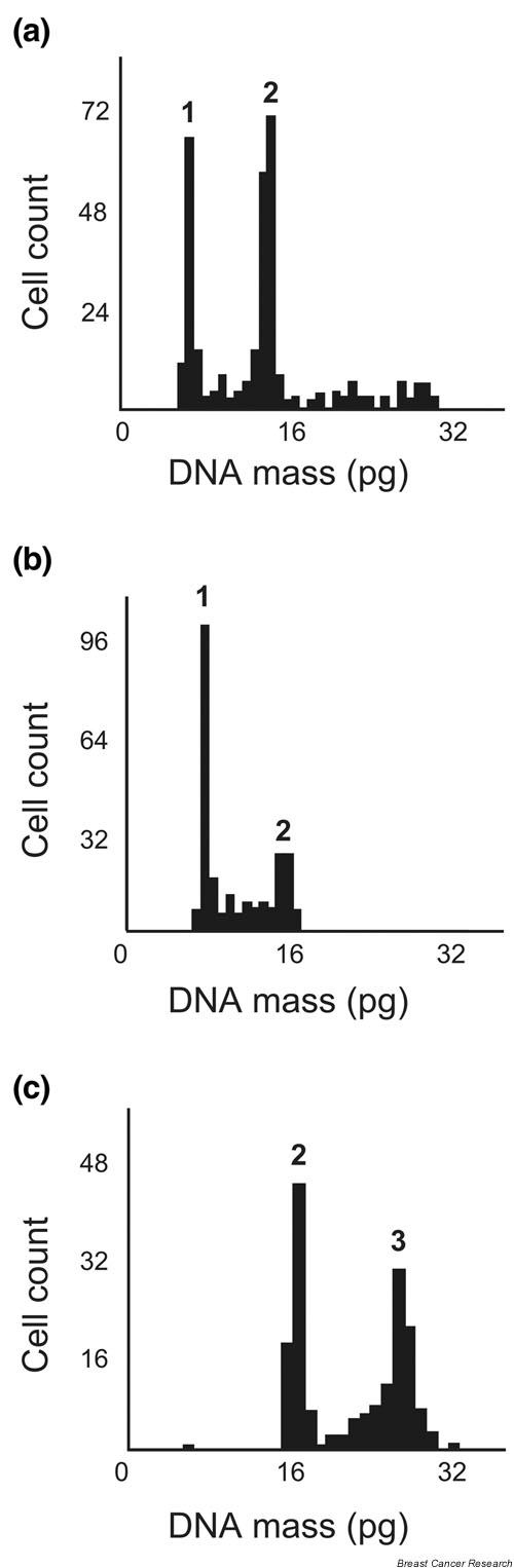
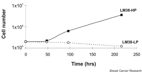
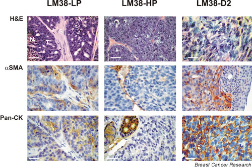
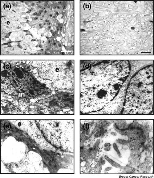
Methods: To characterize this tumor further, primary M38 cultures were established. The low-passage LM38-LP subline contained two main cell components up to the 30th subculture, whereas the higher passage LM38-HP subline was mainly composed of small spindle-shaped cells. In addition, a large spindle cell clone (LM38-D2) was established by dilutional cloning of the low-passage MM38-LP cells. These cell lines were studied by immunocytochemistry, electron microscopy and ploidy, and syngeneic mice were inoculated subcutaneously and intravenously with the different cell lines, either singly or combined to establish their tumorigenic and metastatic capacity.

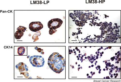
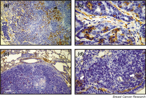
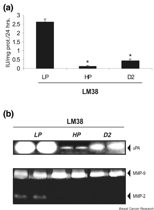
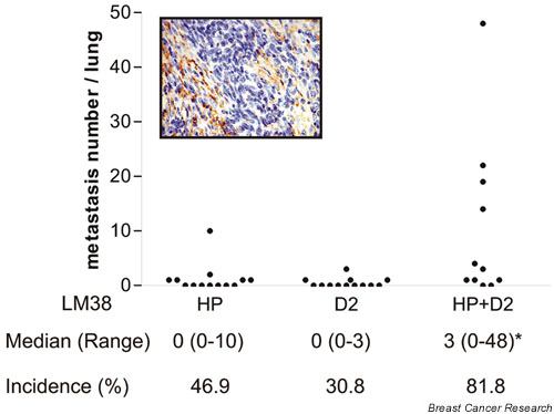
Results: The two subpopulations of LM38-LP cultures were characterized as luminal and myoepithelium-like cells, whereas LM38-HP was mainly composed of small, spindle-shaped epithelial cells and LM38-D2 contained only large myoepithelial cells. All of them were tumorigenic when inoculated into syngeneic mice, but only LM38-LP cultures containing both conserved luminal and myoepithelial malignant cells developed aggressive papillary adenocarcinomas that spread to lung and regional lymph nodes.

Conclusion: The differentiated histopathology and metastatic ability of the spontaneous transplantable M38 murine mammary tumor is associated with the presence and/or interaction of both luminal and myoepithelial tumor cell types.


Figures

Figure 1
Figure 1
Immunocytochemical characterization of LM38-LP, LM38-HP, and LM38-D2 cells (top, middle, and bottom panels respectively). Cell monolayers were stained for the expression of pancytokeratins (pan-CK; left panels) and α-smooth muscle actin (α-SMA; right panels). The LM38-LP cell subline consisted mainly of islets of epithelial cells intensely positive for pan-CK surrounded by α-SMA-positive large myoepithelial cells. The LM38-HP subline predominantly showed small spindle cells positive for pan-CK, which do not form islets, and sparse α-SMA-positive altered myoepithelial cells. The LM38-D2 clone was homogenously made up of α-SMA-stained myoepithelial cells, also positive for pan-CK. (Original magnification ×400; scale bar, 35 μm.)
Figure 2
Figure 2
Dual staining for E-cadherin (upper panels) and actin (lower panels) of LM38-LP, LM38-HP, and LM38-D2 cells (left, middle, and right panels, respectively). Whereas islets of epithelial cells showed evident E-cadherin staining of membranes, neither myoepithelial cells (in LM38-LP or LM38-D2 cultures) nor LM38-HP epithelioid cells did so. Actin was distributed cortically in the islets of epithelial cells, formed long stress fibres in the myoepithelial cells, and showed poor cortical distribution and the simultaneous presence of stress fibers in the spindle-shaped LM38-HP cell line. (Original magnification ×400; scale bar, 35 μm.)
Figure 3
Figure 3
Analysis of DNA ploidy of LM38-LP (a), LM38-HP (b) and LM38-D2 (c) cells. (a) Whereas islets of epithelial cells were hypotriploid (1), large spindle cells, probably of myoepithelial phenotype, made up a large hypertetraploid subpopulation (2). (b) The main small spindle cell subpopulation was hypotriploid (1), similarly to the islets of epithelial cells in LM38-LP cultures. A small peak of a hypertetraploid subpopulation was also found (2). (c) The myoepithelial LM38-D2 clone was made up of two subpopulations, either hypertetraploid (2) or even with a higher DNA index (3).
Figure 4
Figure 4
Morphology and cellular composition of spheroids formed in a three-dimensional growth assay. LM38-LP cells (left panels) formed glandular-like smooth-bordered spheroids, whereas LM38-HP cells (right panels) grew in loose irregular clusters. Cells positive for pancytokeratins (pan-CK; upper panels) formed a polarized epithelial layer surrounding a kind of lumen in LM38-LP spheroids, but were disorganized in LM38-HP clusters. In LM38-LP spheroids myoepithelial cells, stained for either cytokeratin 14 (CK14) or α-smooth muscle actin (α-SMA; lower panels), were found as a basal cell layer (arrow) surrounding luminal epithelial cells or occupying the spheroid core in an inside-out display (arrowhead). Some CK14-positive myoepithelial cells were found intermingled in LM38-HP clusters (arrow). Inset: Double staining of LM38-LP spheroids: α-SMA (light brown), pan-CK (blue), nuclei (red). (Original magnification ×400; scale bar, 35 μm.)
Figure 5
Figure 5
Growth curves of LM38 sublines in a three-dimensional assay in liquid medium. Cell suspensions (2 × 10⁵ cells/ml) in complete growth medium were seeded on top of an agar layer. At different time points spheroids were collected and trypsin-treated; cells were then counted. Whereas the LM38-HP cells proliferated at least up to the 10th day in this condition, the LM38-LP subline not only did not proliferate but began to die after the fourth day. The representative data shown correspond to one of two independent experiments. Standard deviations (not shown) were within ± 10% of the mean.
Figure 6
Figure 6
Histopathology of subcutaneous tumors formed after the inoculation of LM38 sublines into syngeneic mice. Tumor sections were stained with hematoxylin and eosin (upper panels) or by an immunohistochemistry procedure, for α-smooth muscle actin (α-SMA; middle panels), for pancytokeratins (pan-CK; left and central lower panels) or for cytokeratin 14 (CK14; right lower panel). The LM38-LP cells (left panels) formed differentiated papillary adenocarcinomas made up of cells positive for α-SMA and pan-CK surrounding fibrovascular strands. In contrast, the LM38-HP cells (central panels) formed poorly differentiated adenocarcinomas with no evidence of glandular structures, that were made up of cells mostly negative for both cell lineage markers. The LM38-D2 clone (right panels) grew as an undifferentiated tumor consisting of large cells positive for CK14 and α-SMA. (Original magnification ×400; scale bar, 35 μm.) E, epidermis; F, hair follicle; N, necrosis; S, fibrovascular stroma; T, tumor tissue.
Figure 7
Figure 7
Immunohistochemical staining for α-smooth muscle actin (α-SMA) expression in LM38-LP lymph node (a, b) and lung metastases (c, d). Whereas the lymph node metastasis showed the same histoarchitecture as the subcutaneous primary tumor, and the presence of many α-SMA-positive myoepithelial cells, lung metastatic nodes were less differentiated and only a few α-SMA positive cells could be found (arrow). Scale bars, 140 μm (a,c) and 35 μm (b,d).
Figure 8
Figure 8
Morphological and ultrastructural features of LM38-LP and LM38-HP subcutaneous tumors. Light microscopy of Maraglass-embedded sections confirmed that LM38-LP (a) heterogenous tumor parenchyma was composed of two main cell types: small epithelial cells (e) of light cytoplasm and euchromatic nuclei with distinct nucleoli and some dense chromatin granules found close to the lumen, and basal myoepithelial (m) dark cells with intensely stained irregular nuclei and large nucleoli, placed near the stroma (s) or mixed at random with the small light cells. The LM38-HP tumors (b) were mainly made up of small light epithelial cells. (Original magnification ×400; scale bar,35 μm.) Electron micrographs of LM38-LP tumor showing the two subpopulations at low magnification (c) (original magnification ×8000), a detail of the characteristic nuclei, nucleoli and cytoplasm of the light epithelial cells (d) (original magnification ×14,000) and the presence of abundant parallel microfilament bundles (arrow) and focal densities (arrowhead) in a cytoplasmic process of a dark myoepithelial cell (e) (original magnification ×17,000). Glandular lumina with microvilli and rudimentary intercellular junctions in LM38-HP tumor (f) (original magnification ×40,000), confirming its glandular epithelial origin.
Figure 9
Figure 9
Secretion of proteases in vitro by the LM38 sublines. (a) Quantification of secreted urokinase-type plasminogen activator (uPA) activity by radial caseinolysis. The LM38-HP cells and LM38-D2 clone secreted significantly less uPA than LM38-LP cells. (b) Upper panel: zymogram of media conditioned by LM38-LP, LM38-HP, and LM38-D2 cells showing a main 48 kDa uPA band, confirming the differential activity observed in the radial assay. Lower panel: gelatin co-polymerized zymogram showing that only the highly metastatic LM38-LP cells were able to secrete matrix metalloproteinase (MMP)-2 to the culture medium. *P < 0.05 compared with LM38-LP.
Figure 10
Figure 10
Co-inoculation experiments in vivo. LM38-HP and LM38-D2 cells were cultivated together before subcutaneous injection, as described in the Materials and methods section. The combined tumors showed a higher incidence and number of spontaneous lung metastases than LM38-HP or LM38-D2 tumors. The results of two separate experiments are presented together. *P < 0.05 compared with LM38-HP and LM38-D2. Inset: a representative subcutaneous combined tumor stained for α-smooth muscle actin. Although myoepithelial cells surround epithelial cell nests, the characteristic papillary organization of M38 and LM38-LP tumors is not found.

Similar articles

Cited by

References

    1. Lakhani S, O'Hare M. The mammary epithelial cell – Cinderella or ugly sister? Breast Cancer Res. 2001;3:1–4. doi: 10.1186/bcr260. - DOI - PMC - PubMed
    1. Deugnier MA, Teulière J, Faraldo M, Thiery JP, Glukhova MA. The importance of being a myoepithelial cell. Breast Cancer Res. 2002;4:224–230. doi: 10.1186/bcr459. - DOI - PMC - PubMed
    1. Liu QY, Niranjan B, Gomes P, Gomm J, Davies D, Coombes C, Buluwela L. Inhibitory effects of activin on the growth and morphogenesis of primary and transformed mammary epithelial cells. Cancer Res. 1996;56:1155–1163. - PubMed
    1. Rudland PS, Fernig DG, Smith JA. Growth factors and their receptors in neoplastic mammary glands. Biomed Pharmacother. 1995;49:389–399. doi: 10.1016/0753-3322(96)82676-X. - DOI - PubMed
    1. Xiao G, Liu YE, Gentz R, sang QA, Ni J, Goldberg ID, Shi YE. Suppression of breast cancer growth and metastasis by a serpin myoepithelium derived serine protease inhibitor expressed in the mammary myoepithelial cells. Proc Natl Acad Sci. 1999;96:3700–3705. doi: 10.1073/pnas.96.7.3700. - DOI - PMC - PubMed

Publication types

MeSH terms