. 2004;6(1):28-38.
doi: 10.1186/ar1155. Epub 2004 Feb 5.

Flow cytometric assessment of the signaling status of human B lymphocytes from normal and autoimmune individuals

Affiliations

Flow cytometric assessment of the signaling status of human B lymphocytes from normal and autoimmune individuals

Amrie C Grammer et al. Arthritis Res Ther. 2004.

Abstract

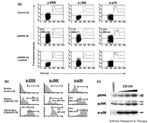
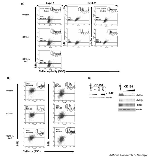
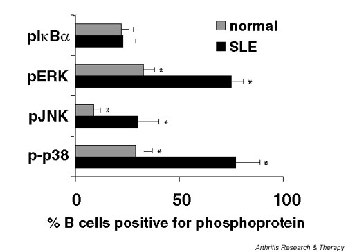
Abnormalities in lymphocyte signaling cascades are thought to play an important role in the development of autoimmune disease. However, the large amount of cellular material needed for standard biochemical assessment of signaling status has made it difficult to evaluate putative abnormalities completely using primary lymphocytes. The development of technology to employ intracellular staining and flow cytometry to assess the signaling status of individual cells has now made it possible to delineate the perturbations that are present in lymphocytes from patients with autoimmune disease. As an example, human B cells from the Ramos B cell line and the periphery of systemic lupus erythematosus (SLE) patients or normal nonautoimmune controls were assessed for activation of the NF-kappaB and mitogen activated protein kinase (MAPK) signaling cascades by intracellular multiparameter flow cytometric analysis and biochemical Western blotting. In combination with fluorochrome conjugated antibodies specific for surface proteins that define B cell subsets, antibodies that recognize activated, or phosphorylated inhibitors of kappaB (IkappaB) as well as the extracellular regulated kinase (ERK), jun N-terminal kinase (JNK) or p38 MAPKs were used to stain fixed and permeabilized human B cells and analyze them flow cytometrically. Examination of the known signaling pathways following engagement of CD40 on human B cells confirmed that intracellular flow cytometry and Western blotting equivalently assay CD154-induced phosphorylation and degradation of IkappaB proteins as well as phosphorylation of the MAPKs ERK, JNK and p38. In addition, B cells from the periphery of SLE patients had a more activated status immediately ex vivo as assessed by intracellular flow cytometric analysis of phosphorylated ERK, JNK and p38 when compared with B cells from the periphery of normal, nonautoimmune individuals. Together, these results indicate that multiparameter intracellular flow cytometric analysis of signaling pathways, such as the NF-kappaB and MAPK cascades, can be used routinely to assess the activation status of a small number of cells and thus delineate abnormalities in signaling molecules expressed in primary lymphocytes from patients with autoimmune disease.


Figures

Figure 1
Figure 1
Assessment of extracellular regulated kinase (ERK), jun N-terminal kinase (JNK) and p38 mitogen activated protein kinase (MAPK) activity in human B lymphocytes by intracellular flow cytometric analysis. R2G6 cells (3 × 10⁵) were fixed, permeabilized, incubated in the presence or absence of a blocking peptide and stained with isotype matched control antibodies or phosphospecific antibodies that recognize pERK, pJNK and p-p38 (a) immediately or (b) following incubation with increasing amounts of rCD154 for 15 min at 37°C. Dot plots of cell size on the x-axis versus fluorescence intensity in the channel that detects the fluorochrome conjugated secondary antibody on the y-axis are shown in (a). Histograms of fluorescence intensity of the channel that detects the fluorochrome conjugated secondary antibody are shown in (a) and (b). The percentage of positive cells and the mean fluorescence intensity (MFI) of positive staining are indicated. The solid line indicates the division between negative background staining of isotype-matched control antibody and positive staining with the pMAPK antibody. (c) Western blot analysis of pERK, pJNK or p-p38 expression in R2G6 cells following incubation with rCD154 for 15 min at 37°C.
Figure 2
Figure 2
Assessment of NF-κB activation in human B lymphocytes by multiparameter intracellular flow cytometric analysis of IκB isoforms (-α, -β, -ε) and the phosphorylation status of IκBα. R2G6 cells (3 × 10⁵) were incubated with rCD154 in the presence or absence of 30 μM of the proteosome inhibitor lactacystein (LAC) or the inhibitor of IκB phosphorylation BAY11-7082 (BAY; Calbiochem) for 15 min at 37°C. Cells were fixed, permeabilized and stained with isotype-matched control antibody or antibody that recognize (a) IκBα, pIκBα, (b) IκBβ or IκBε. Two independent experiments are presented in both (a) and (b). Experiment 1 in (a) depicts IκBα staining following CD154 stimulation in the presence or absence of LAC. Experiment 2 in (b) depicts IκBα and pIκBα staining in the presence or absence of LAC. It is important to note that R2G6 cells are known to have a high level of constitutive NF-κB activation, accounting for the large percentage of cells expressing p-IκBα. Experiment 1 in (b) depicts staining for IκBβ following stimulation with CD154 in the presence or absence of LAC. Experiment 2 in (b) depicts staining for IκBε following stimulation with CD154 in the presence or absence of LAC. Dot plots of cellular size or complexity on the x-axis versus fluorescence intensity in the channel that detects the fluorochrome conjugated secondary antibody on the y-axis are shown in (a) and (b). Insets of histograms of fluorescence intensity of the channel that detects the fluorochrome conjugated secondary antibody are shown in (a) and (b). The percentage of positive cells and the mean fluorescence intensity (MFI) of positive staining are indicated. The solid line indicates the division between negative background staining of isotype-matched control antibody and positive staining with the antibody that recognizes a specific IκB. (c) Western blot analysis of pIκBα as well as total IκBα, -β and -ε following CD154 stimulation in the presence or absence of LAC or BAY11-7082 (BAY).
Figure 3
Figure 3
Multiparameter intracellular flow cytometry reveals elevated percentages of B lymphocytes in the periphery of systemic lupus erythematosus (SLE) patients with spontaneous activation of extracellular regulated kinase (ERK), jun N-terminal kinase (JNK) and p38 mitogen activated protein kinases (MAPKs). Peripheral B lymphocytes isolated from SLE patients (n = 7) or nonautoimmune normal individuals (n = 7) were fixed, permeabilized and stained with phosphospecific antibody for pIκBα, pERK, pJNK or p-p38. The mean ± SEM percentages of CD19+ B cells positive for each phospho-Ab are shown graphically. *P < 0.05 by Student's t test.

References

    1. Far DF, Peyron JF, Imbert V, Rossi B. Immunofluorescent quantification of tyrosine phosphorylation of cellular proteins in whole cells by flow cytometry. Cytometry. 1994;15:327–334. - PubMed
    1. Far DF, Rossi B. Measurement of protein tyrosine phosphorylation in T-cell subsets by flow cytometry. Methods Mol Biol. 2000;134:301–306. - PubMed
    1. Vuillier F, Scott-Algara D, Cayota A, Siciliano J, Nugeyre MT, Dighiero G. Flow cytometric analysis of protein-tyrosine phosphorylation in peripheral T cell subsets. Application to healthy and HIV-seropositive subjects. J Immunol Methods. 1995;85:43–56. doi: 10.1016/0022-1759(95)00103-H. - DOI - PubMed
    1. Hubert P, Grenot P, Autran B, Debre P. Analysis by flow cytometry of tyrosine-phosphorylated proteins in activated T-cell subsets on whole blood samples. Cytometry. 1997;29:83–91. doi: 10.1002/(SICI)1097-0320(19970901)29:1<83::AID-CYTO9>3.3.CO;2-3. - DOI - PubMed
    1. Perez OD, Nolan GP. Simultaneous measurement of multiple active kinase states using polychromatic flow cytometry. Nat Biotechnol. 2002;20:155–162. - PubMed