. 2004;6(1):R46-R55.
doi: 10.1186/ar1024. Epub 2003 Nov 12.

Does protein kinase R mediate TNF-alpha- and ceramide-induced increases in expression and activation of matrix metalloproteinases in articular cartilage by a novel mechanism?

Affiliations

Does protein kinase R mediate TNF-alpha- and ceramide-induced increases in expression and activation of matrix metalloproteinases in articular cartilage by a novel mechanism?

Sophie J Gilbert et al. Arthritis Res Ther. 2004.

Abstract

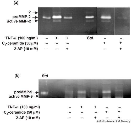
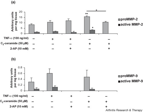
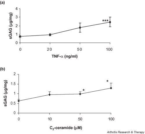
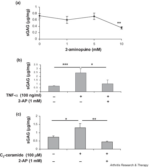
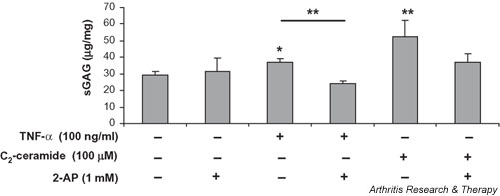
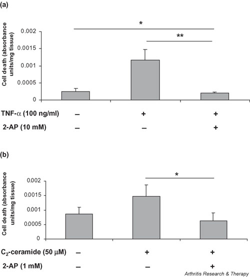
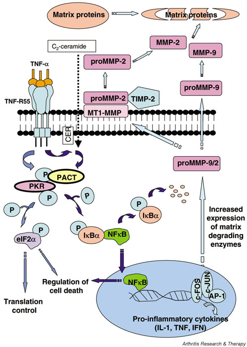
We investigated the role of the proinflammatory cytokine TNF-alpha, the second messenger C2-ceramide, and protein kinase R (PKR) in bovine articular cartilage degradation. Bovine articular cartilage explants were stimulated with C2-ceramide or TNF-alpha for 24 hours. To inhibit the activation of PKR, 2-aminopurine was added to duplicate cultures. Matrix metalloproteinase (MMP) expression and activation in the medium were analysed by gelatin zymography, proteoglycan release by the dimethylmethylene blue assay, and cell viability by the Cytotox 96(R) assay. C2-ceramide treatment of cartilage explants resulted in a significant release of both pro- and active MMP-2 into the medium. Small increases were also seen with TNF-alpha treatment. Incubation of explants with 2-aminopurine before TNF-alpha or C2-ceramide treatment resulted in a marked reduction in expression and activation of both MMP-2 and MMP-9. TNF-alpha and C2-ceramide significantly increased proteoglycan release into the medium, which was also inhibited by cotreatment with 2-aminopurine. A loss of cell viability was observed when explants were treated with TNF-alpha and C2-ceramide, which was found to be regulated by PKR. We have shown that C2-ceramide and TNF-alpha treatment of articular cartilage result in the increased synthesis and activation of MMPs, increased release of proteoglycan, and increased cell death. These effects are abrogated by treatment with the PKR inhibitor 2-aminopurine. Collectively, these results suggest a novel role for PKR in the synthesis and activation of MMPs and support our hypothesis that PKR and its activator, PACT, are implicated in the cartilage degradation that occurs in arthritic disease.


Figures

Figure 1
Figure 1
Detection of (a) MMP-2 and (b) MMP-9 activity in media collected from bovine articular cartilage explants treated with TNF-α or C2-ceramide for 24 hours. Medium was collected 24 hours after treatment of explants with 100 ng/ml TNF-α or 50 μM C2-ceramide in the presence or absence of the PKR inhibitor 2-AP (10 mM) and analysed by gelatin substrate zymography. A standard known to contain either MMP-2 or MMP-9 was included (Std). An unidentified enzyme is indicated by '?'. 2-AP, 2-aminopurine; MMP, matrix metalloproteinase; TNF, tumour necrosis factor.
Figure 2
Figure 2
Quantitative analysis of (a) MMP-2 and (b)MMP-9 synthesis by bovine articular cartilage. Medium was collected 24 hours after treatment of explants with 100 ng/ml TNF-α or 50 μM C2-ceramide in the presence or absence of 2-AP (10 mM) and analysed by gelatin substrate zymography. The area (absorbance units) of substrate gel cleared by proMMP-9 and active MMP-9 was measured by scanning densitometry. Each area obtained was related to the gel clearance obtained with the fibroblast-conditioned-medium standard in order to facilitate comparisons between gels. Data shown are arbitrary units per milligram of wet weight of tissue and are expressed as means ± SEM. *Significantly different from control explants at P < 0.05; **P < 0.01. 2-AP, 2-aminopurine; MMP, matrix metalloproteinase; TNF, tumour necrosis factor.
Figure 3
Figure 3
Treatment with TNF-α or ceramide induces proteoglycan release from articular cartilage. Cartilage explants were cultured for 24 hours in the presence of (a) TNF-α (0–100 ng/ml) or (b) C2-ceramide (0–100 μM) and media were analysed for release of sulfated GAGs by dimethylmethylene blue assay. Differences in the release of sGAG associated with culture treatment are expressed as micrograms GAG per milligram wet weight of cartilage. *Significantly different from untreated, control explants at P < 0.05; **P < 0.001. sGAG, sulfated glycosaminoglycan; TNF, tumour necrosis factor.
Figure 4
Figure 4
The PKR inhibitor 2-AP blocks both basal and TNF-α and C2-ceramide-induced proteoglycan release from articular cartilage. Bovine articular cartilage explants were cultured for 24 hours in the presence of (a) various concentrations of 2-AP (0–10 mM) alone, (b) TNF-α (100 ng/ml) with or without 2-AP (1 mM) or (c) C2-ceramide (100 μM) with or without 2-AP (1 mM). Medium was analysed for release of sGAGs by dimethylmethylene blue assay, expressed as micrograms of glycosaminoglycan released per milligram wet weight of cartilage. *Significantly different from control explants at P < 0.05; **P < 0.01; ***P < 0.001. 2-AP, 2-aminopurine; PKR, protein kinase R; sGAG, sulfated glycosaminoglycan; TNF, tumour necrosis factor.
Figure 5
Figure 5
The PKR inhibitor 2-AP blocks TNF-α and C2-ceramide-induced proteoglycan synthesis. Cartilage explants were cultured for 24 hours in the presence of 2-AP (1 mM) alone, TNF-α (100 ng/ml) with or without 2-AP (1 mM) or C2-ceramide (100 μM) with or without 2-AP (1 mM). Explants were digested by papain (300 μg/ml) as described in Materials and methods and the amount of sGAG present determined as described above and expressed as micrograms glycosaminoglycan per milligram wet weight of cartilage. *Significantly different from control explants at P < 0.05; ** P < 0.01. 2-AP, 2-aminopurine; PKR, protein kinase R; sGAG, sulfated glycosaminoglycan; TNF, tumour necrosis factor.
Figure 6
Figure 6
TNF-α and C2-ceramide induce cell death in articular cartilage via a mechanism involving PKR. Bovine articular cartilage explants were cultured for 24 hours in the presence of (a)TNF-α (100 ng/ml) or (b) C2-ceramide (50 μM). 2-AP (1 or 10 mM) was added 1 hour before the addition of treatments. Viability of explant tissue was determined using the CytoTox 96® assay, which quantitatively measures lactate dehydrogenase released into culture media upon cell death during the culture period. Data shown are absorbance units per milligrams of starting tissue and are expressed as means ± SEM. *Significantly different from control explants at P < 0.05; **P < 0.01. 2-AP, 2-aminopurine; PKR, protein kinase R; TNF, tumour necrosis factor.
Figure 7
Figure 7
The potential role of PKR in cartilage degradation. Results from the current study have led us to hypothesise that TNF-α-induced degradative pathways in cartilage may be mediated via activation of PKR. TNF-α binding to its receptor (TNF-R55) may activate PKR either directly or through ceramide (CER) to increase transcription and activation of MMPs via induction of NFκB and early response genes (c-fos and c-jun). Activation of PKR and subsequent phosphorylation of eIF2α would lead to an inhibition of protein synthesis and increased apoptosis, which may also affect cartilage integrity. Increased expression and activation of MMPs, in the absence of a corresponding increase in their inhibitors, would shift the balance of homeostasis towards matrix catabolism. AP-1, activator protein-1; eIF2α, eukaryotic initiation factor 2α ; IκBα, inhibitor kappa B alpha; MMP, matrix metalloproteinase; NFκB, nuclear factor κB; P, phosphorylated; PACT, PKR-activating protein; PKR, protein kinase R; TIMP, tissue inhibitors of MMP; TNF, tumour necrosis factor; TNF-R55, tumour necrosis factor receptor-55.

References

    1. Dean DD, Martel-Pelletier J, Pelletier JP, Howell DS, Woessner JF., Jr Evidence for metalloproteinase and metalloproteinase inhibitor imbalance in human osteoarthritic cartilage. J Clin Invest. 1989;84:678–685. - PMC - PubMed
    1. Aigner T, Zien A, Gehrsitz A, Gebhard PM, McKenna L. Anabolic and catabolic gene expression pattern analysis in normal versus osteoarthritic cartilage using complementary DNA-array technology. Arthritis Rheum. 2001;44:2777–2789. doi: 10.1002/1529-0131(200112)44:12<2777::AID-ART465>3.0.CO;2-H. - DOI - PubMed
    1. Yoshihara Y, Nakamura H, Obata K, Yamada H, Hayakawa T, Fujikawa K, Okada Y. Matrix metalloproteinases and tissue inhibitors of metalloproteinases in synovial fluids from patients with rheumatoid arthritis or osteoarthritis. Ann Rheum Dis. 2000;59:455–461. doi: 10.1136/ard.59.6.455. - DOI - PMC - PubMed
    1. Goldring MB. Osteoarthritis and cartilage: The role of cytokines. Curr Rheumatol Rep. 2000;2:459–465. - PubMed
    1. Westacott CI, Barakat AF, Wood L, Perry MJ, Neison P, Bisbinds I, Armstrong L, Millar AB, Elson CJ. Tumour necrosis factor-alpha can contribute to focal loss of cartilage in osteoarthritis. Osteoarthritis Cartilage. 2000;8:213–221. doi: 10.1053/joca.1999.0292. - DOI - PubMed

LinkOut - more resources