Comparative Study
. 2004 Feb 20;319(2):249-61.
doi: 10.1016/j.virol.2003.10.036.

Apoptosis in astrovirus-infected CaCo-2 cells

Affiliations
Comparative Study

Apoptosis in astrovirus-infected CaCo-2 cells

Susana Guix et al. Virology. .

Abstract

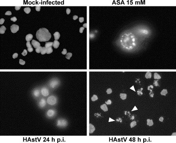
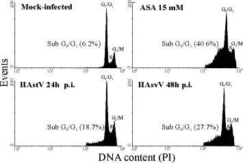
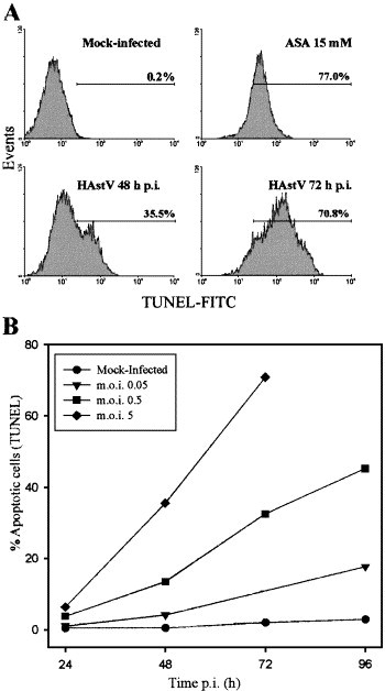
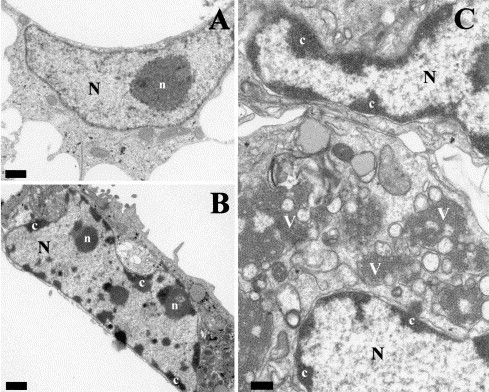
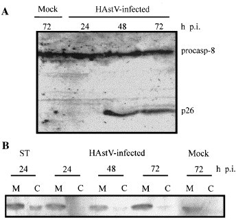
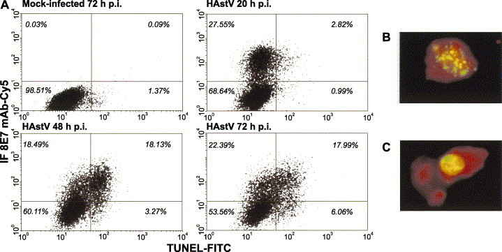
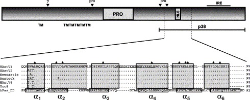
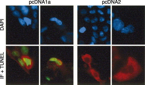
Cell death processes during human astrovirus replication in CaCo-2 cells and their underlying mechanisms were investigated. Morphological and biochemical alterations typical of apoptosis were analyzed in infected cells using a combination of techniques, including DAPI staining, the sub-G(0)/G(1) technique and the TUNEL assay. The onset of apoptosis was directly proportional to the virus multiplicity of infection. Transient expression experiments showed a direct link between astrovirus ORF1a encoded proteins and apoptosis induction. A computer analysis of the astrovirus genome revealed the presence of a death domain in the nonstructural protein p38 of unknown function, encoded in ORF1a. Apoptosis inhibition experiments suggested the involvement of caspase 8 in the apoptotic response, and led to a reduction in the infectivity of the virus progeny released to the supernatant. We conclude that apoptotic death of host cells seems necessary for efficient human astrovirus replication and particle maturation.


Figures

Fig. 1
Fig. 1
Morphology of cell nuclei of mock-infected CaCo-2 cells, aspirin-treated cells (ASA 15 mM) and HAstV-infected cells at 24 and 48 h p.i., after DNA staining with DAPI. At 48 h p.i., many infected cells show apoptotic bodies due to the condensation of chromatin in several micronuclei (arrows).
Fig. 2
Fig. 2
Flow cytometric analysis of DNA content of propidium iodide (PI)-stained CaCo-2 cells. The percentage of apoptotic cells was obtained by calculating the percentage of the cell population showing a DNA content lower than G0/G1 cells in the cell cycle.
Fig. 3
Fig. 3
In situ detection of apoptosis by TUNEL analysis. (A) Flow cytometry analysis of mock-infected cells at 48 h p.i., aspirin-treated cells at 48 h and HAstV-infected cells at 48 and 72 h p.i. after TUNEL staining of apoptotic nuclei. The percentage of apoptotic cells was estimated as the percentage of cells showing FITC fluorescence nuclei, due to the addition of fluorescein dUTP at 3′OH ends of fragmented DNA. A cursor for FITC positivity was defined from mock-infected cultures, which usually included 0.1–3.0% of the total cell population (in the depicted experiment, it includes only 0.2% of the total population of mock-infected cells). (B) Dose-dependent and time-dependent appearance of apoptotic cells. Cells were infected with different m.o.i., harvested at various times p.i., analyzed by TUNEL assay and subjected to flow cytometry. The values of the error bars, which are not depicted for the sake of clarity, were always below 0.44× the mean value.
Fig. 4
Fig. 4
Electron micrographs of mock-infected (A) and HAstV-infected CaCo-2 cells at 48 h p.i. (B and C). In mock-infected cells, the large nucleus (N) displays a large, unique nucleolus (n). HAstV-infected cells are characterized by numerous masses of condensed chromatin (c) dispersed at the periphery of a convoluted nucleus. Aggregates of astrovirus particles (v) accumulated in the cytoplasm of infected cells (C). Bars equal 1 μm in A and B, and 0.5 μm in C.
Fig. 5
Fig. 5
FLICE/caspase-8 cleavage and distribution of mitochondrial cytochrome c during astrovirus infection. (A) Detection of procaspase-8 and activation-resulting product of prodomain p26 by Western blot analysis at the indicated times p.i., according to Medema et al. (1997). (B) Distribution of cytochrome c in mitochondrial (M) and cytosolic fractions (C), in 1 μM staurosporine (ST)-treated cells, astrovirus-infected cells at various times p.i. and mock-infected CaCo-2 cells at 72 h p.i.
Fig. 6
Fig. 6
Simultaneous determination of astrovirus antigens and apoptosis by flow cytometry. After apoptosis detection by TUNEL assay, an immunofluorescence analysis was performed on mock-infected and HAstV-infected CaCo-2 cells harvested at different times p.i. (A) Representative diagram of flow cytometry double labeling results. Cutoff values for positive cells were set so that more than 99.5% of total cells belonged to the lower left quadrant, in a dot plot of infected cells that had been labeled with negative control solution provided by the manufacturers for TUNEL and incubated with the cyanine5-conjugated anti-mouse antibody but not the astrovirus-specific monoclonal antibody (not shown). The total percentage of cells belonging to each quadrant is indicated. (B and C) Analysis of morphological changes of HAstV-infected cells at 72 h p.i. under a fluorescence microscope. Red color corresponds to the virus immunofluorescence, while green color corresponds to the FITC labeling of fragmented DNA due to apoptosis.
Fig. 7
Fig. 7
Schematic diagram of astrovirus ORF1a. Predicted transmembrane helices (TM), protease motif (PRO), predicted nuclear localization signal (NLS) and an immunoreactive epitope (IRE) are shown. Black arrowheads refer to putative proteolytic cleavage sites, which seem to be dependent on both cellular proteases (?) and the viral protease (pro). The predicted death domain within ORF1a is indicated and a structure-based sequence alignment of all available HAstV sequences and the amino acid sequence of hFas death domain is shown. Helices are indicated by boxes and conserved hydrophobic core residues are indicated by an asterisk above the sequence. Underlined sequence corresponds to the NLS. Accession numbers L23513 (HAstV1), L13745 (HAstV2), Z25771 (A2/88 Newcastle), AF141381 (Rostock), AY257977 (HAstV4 p23795) and AF260508 (Yuc8).
Fig. 8
Fig. 8
Double labeling analysis of HAstV protein expression and apoptosis after transient expression of plasmids containing the full-length ORF1a and ORF2 constructs. Nuclei of all cells were counterstained with DAPI. Red color corresponds to immunofluorescence labeling and green color corresponds to apoptotic nuclei labeled by TUNEL.

References

    1. Abad F.X., Villena C., Guix S., Caballero S., Pintó R.M., Bosch A. Potential role of fomites in the vehicular transmission of human astroviruses. Appl. Environ. Microbiol. 2001;37:3904–3907. - PMC - PubMed
    1. Al-Molawi N., Beardmore V.A., Carter M.J., Kass G.E.N., Roberts L.O. Caspase-mediated cleavage of the feline calicivirus capsid protein. J. Gen. Virol. 2003;84:1237–1244. - PubMed
    1. Alonso C., Oviedo J.M., Martin-Alonso J.M., Diaz E., Boga J.A., Parra F. Programmed cell death in the pathogenesis of rabbit hemorrhagic disease. Arch. Virol. 1998;143:321–332. - PubMed
    1. Ammendolia M.G., Tinari A., Calcabrini A., Superti F. Poliovirus infection induces apoptosis in CaCo-2 cells. J. Med. Virol. 1999;59:122–129. - PubMed
    1. An S., Chen C., Yu X., Leibowitz J.L., Makino S. Induction of apoptosis in murine coronavirus-infected cultured cells and demonstration of E protein as an apoptosis inducer. J. Virol. 1999;73:7853–7859. - PMC - PubMed

Publication types

LinkOut - more resources