. 2004 Mar 1;164(5):689-99.
doi: 10.1083/jcb.200312028. Epub 2004 Feb 23.

Regulation of Ras-MAPK pathway mitogenic activity by restricting nuclear entry of activated MAPK in endoderm differentiation of embryonic carcinoma and stem cells

Affiliations

Regulation of Ras-MAPK pathway mitogenic activity by restricting nuclear entry of activated MAPK in endoderm differentiation of embryonic carcinoma and stem cells

Elizabeth R Smith et al. J Cell Biol. .

Abstract

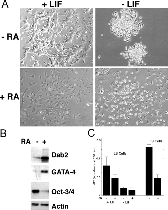
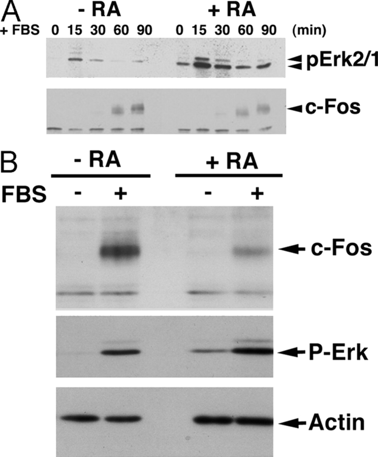
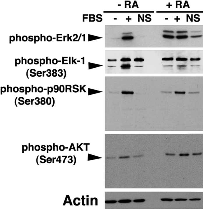
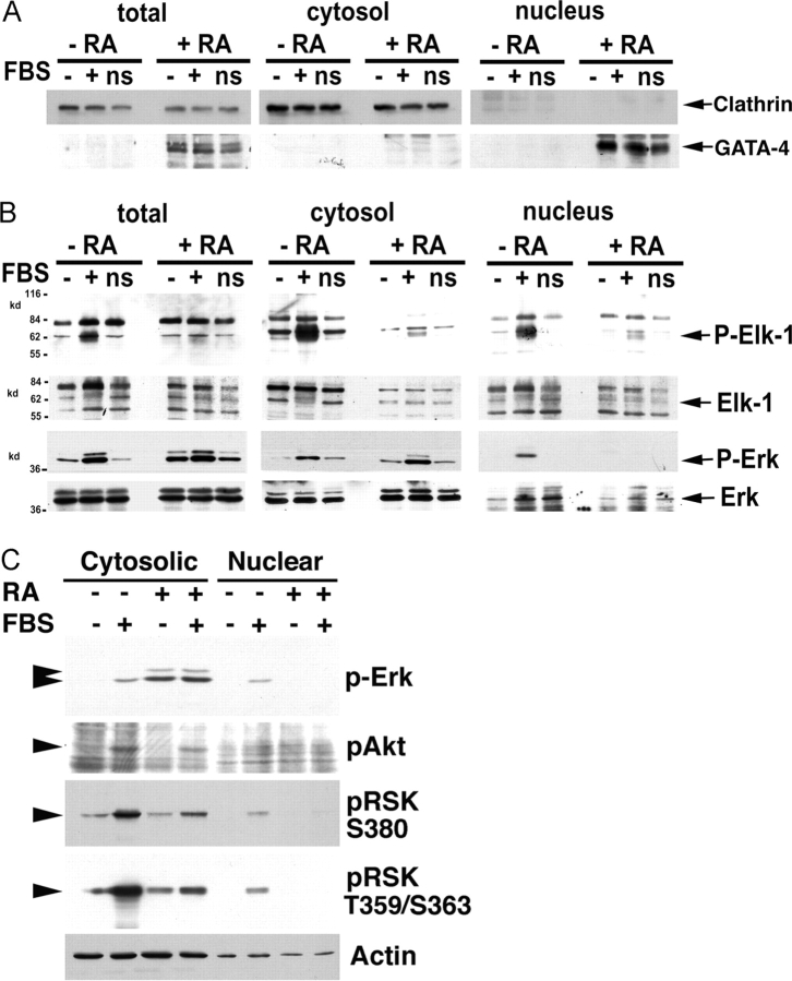
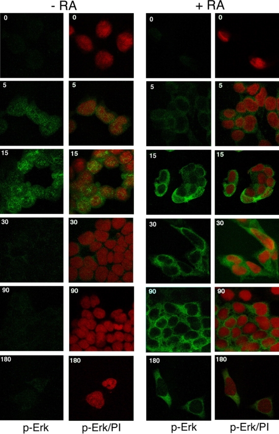
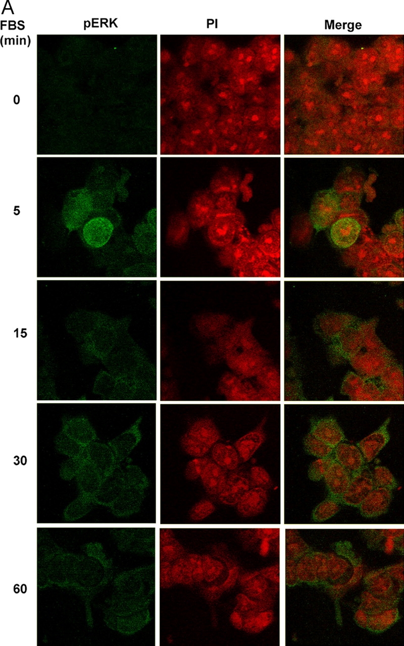
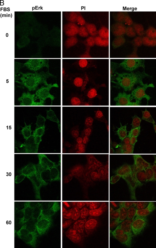
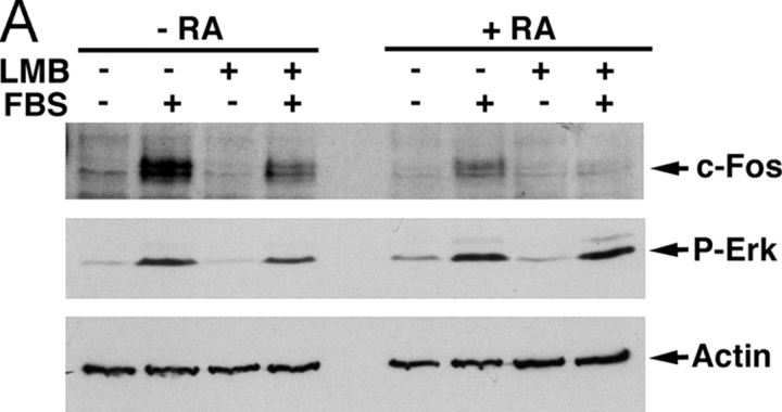
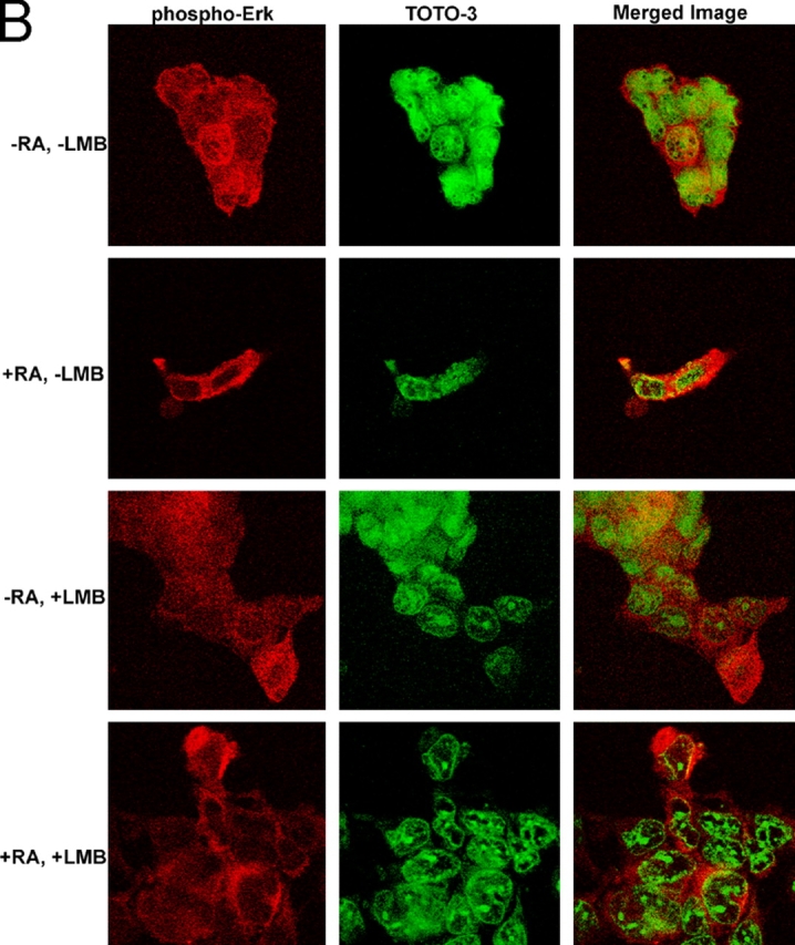
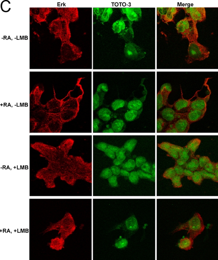
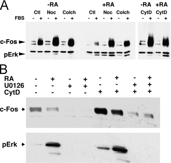
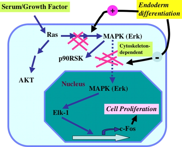
In response to retinoic acid, embryonic stem and carcinoma cells undergo differentiation to embryonic primitive endoderm cells, accompanied by a reduction in cell proliferation. Differentiation does not reduce the activation of cellular MAPK/Erk, but does uncouple mitogen-activated protein kinase (MAPK) activation from phosphorylation/activation of Elk-1 and results in inhibition of c-Fos expression, whereas phosphorylation of the cytoplasmic substrate p90RSK remains unaltered. Cell fractionation and confocal immunofluorescence microscopy demonstrated that activated MAPK is restricted to the cytoplasmic compartment after differentiation. An intact actin and microtubule cytoskeleton appears to be required for the restriction of MAPK nuclear entry induced by retinoic acid treatment because the cytoskeletal disrupting agents nocodazole, colchicine, and cytochalasin D are able to revert the suppression of c-Fos expression. Thus, suppression of cell proliferation after retinoic acid-induced endoderm differentiation of embryonic stem and carcinoma cells is achieved by restricting nuclear entry of activated MAPK, and an intact cytoskeleton is required for the restraint.


Figures

Figure 1.
Figure 1.
RA induces differentiation and cell growth suppression similarly in ES and F9 cells. Monolayer mouse ES or F9 cells on gelatin-coated plates were treated with 0.1 μM RA or DMSO solvent control for 4 d. Medium for ES cells is either with or without LIF. (A) ES cells were photographed. (B) ES cells cultured with or without RA and in the presence of LIF were analyzed by Western blot for Disabled-2 (Dab2), GATA-4, and Oct-3/4. β-actin was measured as a protein loading control. (C) Cell number was determined by MTT assay, measuring absorbance at 570 nm. Equal numbers (104) of ES or F9 cells were treated with or without RA and LIF for 4 d, and the cells were then incubated with MTT reagent for 2 h to measure cellular metabolism. Triplicate wells of cells in 96-well plates were used in each experiment, and the mean ± SD is reported here. The experiment was repeated three times with identical results.
Figure 2.
Figure 2.
RA-induced differentiation of ES cells elevates MAPK activation relative to c-Fos expression. (A) Monolayer ES cells were treated with 0.1 μM RA for 4 d, serum starved overnight, and stimulated the next day with 15% FBS. At indicated time points, cells were disrupted in NP-40 lysis buffer and equal protein was loaded for each sample. Activated MAPK (pErk2/1) and c-Fos were determined by immunoblotting of lysates of undifferentiated (−RA) and differentiated (+RA) ES cells stimulated with 15% FBS for 0–90 min. The experiment was repeated three times with identical results. (B) Parallel experiments using F9 cells are shown for comparison. The results of the 0- and 90-min time points are shown.
Figure 3.
Figure 3.
RA-induced differentiation of F9 cells suppresses phosphorylation of nuclear but not cytoplasmic MAPK substrates. F9 cells were treated with 0.1 μM RA for 4 d, serum starved overnight, and stimulated with or without 15% FBS for 15 min. Another set of cells remained nonsynchronized (NS) by culturing in the presence of serum at all times. Cells were disrupted in lysis buffer, and equal protein was loaded for each sample. The cell lysates were analyzed by immunoblotting for phospho-pErk2/1, -Elk-1, -p90RSK, -AKT, and β-actin (Actin) in undifferentiated (−RA) and differentiated (+RA) F9 cells that had been serum starved (−FBS), serum stimulated (+FBS), or grown continuously in the presence of serum, or nonsynchronized (NS).
Figure 4.
Figure 4.
Activated MAPK is localized in the cytoplasmic fraction of differentiated F9 cells, determined by fractionation. F9 cells, induced to differentiate to endoderm-like cells with 0.1 μM RA for 4 d, were serum starved (−FBS), serum restimulated for 15 min (+FBS), or maintained nonsynchronized (NS). Cells were fractionated to obtain nuclear and cytoplasmic fractions, and equal amounts of sample protein were separated on SDS–polyacrylamide (7.5%) gels and immunoblotted for (A) cytosolic (Clathrin) and nuclear (GATA-4) markers or (B) Elk-1, phospho-Elk-1 (P-Elk-1), MAPK/Erk (Erk), and phospho-MAPK/Erk (P-Erk). (C) The nucleocytoplasmic distribution of pErk, pAKT, p90RSK (S380 and T359/S363), and β-actin is shown. Cell fractionation experiments were done four times with similar conclusions.
Figure 5.
Figure 5.
Immunofluorescence microscopic localization of activated MAPK in F9 cells. F9 cells in monolayer culture, with or without differentiation by RA for 4 d, were serum starved overnight and stimulated with serum. At the indicated times, cells were fixed with ice-cold methanol and stained with an antiactivated/phosphorylated p42/p44 MAPK mAb (p-Erk) and analyzed by immunofluorescence confocal microscopy. Control cells were treated with DMSO vehicle and processed identically. Control (−RA) and differentiated (+RA) F9 cells were serum stimulated for 0–180 min, and phospho-MAPK (Cy-2–labeled; green) is shown. Nuclear PI staining is overlayed (p-Erk/PI) with activated MAPK (pErk). The experiment was performed four times with similar results.
Figure 6.
Figure 6.
Cellular distribution of activated MAPK in ES cells determined by immunofluorescence microscopy. ES cells, induced with RA for 4 d, were serum starved overnight and restimulated with serum. At the indicated time points, cells were fixed with ice-cold methanol, stained with an antiactivated p42/p44 MAPK mAb, and analyzed by immunofluorescence confocal microscopy. Control ES cells with added DMSO vehicle without RA were processed identically. (A) Control (−RA) and (B) differentiated (+RA) ES cells were serum stimulated for 0–60 min, and phospho-MAPK (Alexa 488–labeled; green) and nuclear PI (red) are shown individually and merged. The experiment was performed three times with similar results.
Figure 6.
Figure 6.
Cellular distribution of activated MAPK in ES cells determined by immunofluorescence microscopy. ES cells, induced with RA for 4 d, were serum starved overnight and restimulated with serum. At the indicated time points, cells were fixed with ice-cold methanol, stained with an antiactivated p42/p44 MAPK mAb, and analyzed by immunofluorescence confocal microscopy. Control ES cells with added DMSO vehicle without RA were processed identically. (A) Control (−RA) and (B) differentiated (+RA) ES cells were serum stimulated for 0–60 min, and phospho-MAPK (Alexa 488–labeled; green) and nuclear PI (red) are shown individually and merged. The experiment was performed three times with similar results.
Figure 7.
Figure 7.
Leptomycin B does not reverse the inhibition of c-Fos expression or affect MAPK localization in RA-differentiated F9 cells. (A) Differentiated (+RA) and undifferentiated (−RA) F9 cells were serum starved overnight and preincubated with 10 nM leptomycin B (LMB; final concentration) for 4 h, and then stimulated with 15% FBS for 90 min. Cells were lysed in SDS sample buffer separated on SDS–polyacrylamide (7.5%) gels, c-Fos expression and MAPK activation (pErk) was determined by immunoblotting, and β-actin was used as a control. (B and C) Differentiated (+RA) and undifferentiated (−RA) F9 cells were serum starved overnight and incubated with 10 nM LMB for 30 min before and 10 min during stimulation with 15% FBS as described in Adachi et al. (2000). Activated MAPK (B, phospho-Erk) or total MAPK (C, Erk) were detected by rhodamine red–conjugated secondary antibodies (red) and analyzed by immunofluorescence confocal microscopy. Nuclei were counterstained with TOTO-3 (green).
Figure 7.
Figure 7.
Leptomycin B does not reverse the inhibition of c-Fos expression or affect MAPK localization in RA-differentiated F9 cells. (A) Differentiated (+RA) and undifferentiated (−RA) F9 cells were serum starved overnight and preincubated with 10 nM leptomycin B (LMB; final concentration) for 4 h, and then stimulated with 15% FBS for 90 min. Cells were lysed in SDS sample buffer separated on SDS–polyacrylamide (7.5%) gels, c-Fos expression and MAPK activation (pErk) was determined by immunoblotting, and β-actin was used as a control. (B and C) Differentiated (+RA) and undifferentiated (−RA) F9 cells were serum starved overnight and incubated with 10 nM LMB for 30 min before and 10 min during stimulation with 15% FBS as described in Adachi et al. (2000). Activated MAPK (B, phospho-Erk) or total MAPK (C, Erk) were detected by rhodamine red–conjugated secondary antibodies (red) and analyzed by immunofluorescence confocal microscopy. Nuclei were counterstained with TOTO-3 (green).
Figure 8.
Figure 8.
Cytoskeleton-disrupting agents elevate MAPK activation and c-Fos expression in F9 cells. (A) Undifferentiated F9 cells (−RA) and cells differentiated with RA for 4 d (+RA) were serum starved overnight and pretreated with 10 μM nocodazole (Noc), colchicine (Colch), or cytochalasin D (CytD), and solvent control (Ctl) for 30 min before stimulating with 15% serum for 90 min. Serum-containing medium also contained the inhibitors at the same concentration. Equal protein for each sample was separated on 7.5% PAGE gels, and c-Fos and activated MAPK (pErk) were determined by immunoblotting. In this blot, the membranes were incubated with a mixture of antibodies to c-Fos and pErk to determine both signals simultaneously. Shown is a representative experiment repeated in triplicate. (B) F9 cells, with or without RA treatment, with or without cytochalasin D and/or MEK inhibitor U0126 (10 μM), were first serum starved overnight, and then stimulated with serum for 90 min. The cells were harvested to analyze for c-Fos expression and MAPK activation (pErk) by Western blotting.
Figure 9.
Figure 9.
Model for regulation of nucleocytoplasmic MAPK activity in endoderm differentiation of ES and EC cells. MAPK is a downstream effector of the Ras pathway regulated by mitogen such as serum or growth factors. MAPK phosphorylates the cytoplasmic substrate p90 ribosomal S6 kinase (p90RSK) and the nuclear substrate Elk-1. The nuclear activity of MAPK is the transcription-dependent signal and is required for mitogen-stimulated cell proliferation. Endoderm differentiation of ES and EC cells results in cell cytoskeleton-dependent enhanced activation but also restriction of MAPK nuclear entry and suppression of cell growth.

Similar articles

Cited by

References

    1. Adachi, M., M. Fukuda, and E. Nishida. 1999. Two co-existing mechanisms for nuclear import of MAP kinase: passive diffusion of a monomer and active transport of a dimer. EMBO J. 18:5347–5358. - PMC - PubMed
    1. Adachi, M., M. Fukuda, and E. Nishida. 2000. Nuclear export of MAP kinase (ERK) involves a MAP kinase kinase (MEK)-dependent active transport mechanism. J. Cell Biol. 148:849–856. - PMC - PubMed
    1. Altin, J.G., R. Wetts, K.T. Riabowol, and R.A. Bradshaw. 1992. Testing the in vivo role of protein kinase C and c-fos in neurite outgrowth by microinjection of antibodies into PC12 cells. Mol. Biol. Cell. 3:323–333. - PMC - PubMed
    1. Angel, P., and M. Karin. 1991. The role of Jun, Fos and the AP-1 complex in cell-proliferation and transformation. Biochim. Biophys. Acta. 1072:129–157. - PubMed
    1. Aplin, A.E., S.A. Stewart, R.K. Assoian, and R.L. Juliano. 2001. Integrin-mediated adhesion regulates ERK nuclear translocation and phosphorylation of Elk-1. J. Cell Biol. 153:273–281. - PMC - PubMed

Publication types

MeSH terms