. 2004 Mar 1;199(5):731-6.
doi: 10.1084/jem.20031482. Epub 2004 Feb 23.

Spontaneous development of psoriasis in a new animal model shows an essential role for resident T cells and tumor necrosis factor-alpha

Affiliations

Spontaneous development of psoriasis in a new animal model shows an essential role for resident T cells and tumor necrosis factor-alpha

Onur Boyman et al. J Exp Med. .

Abstract

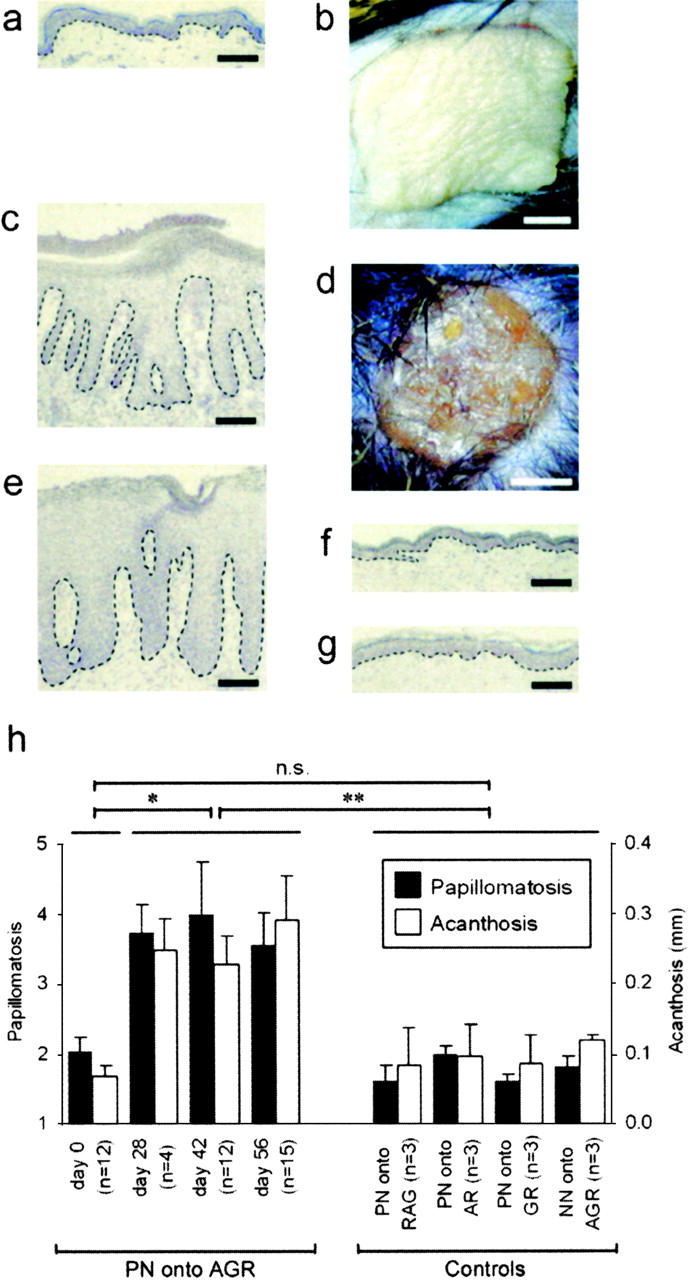
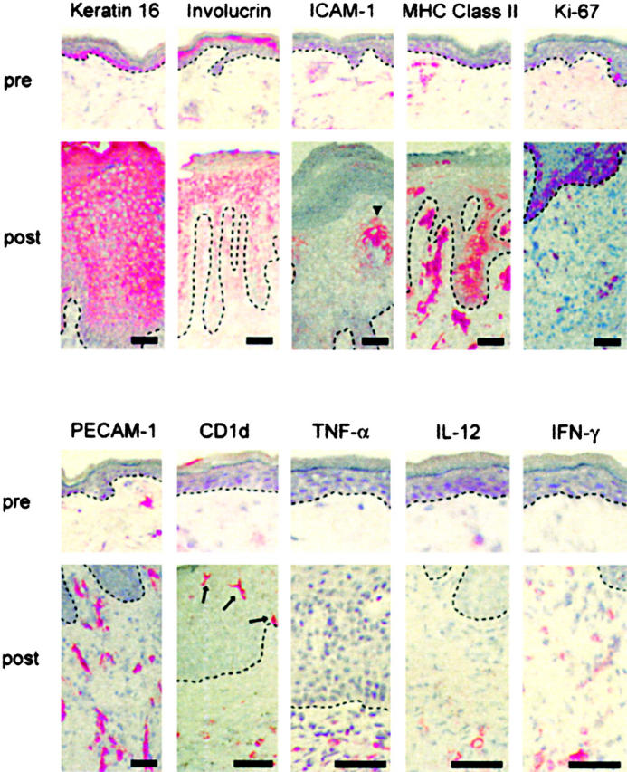
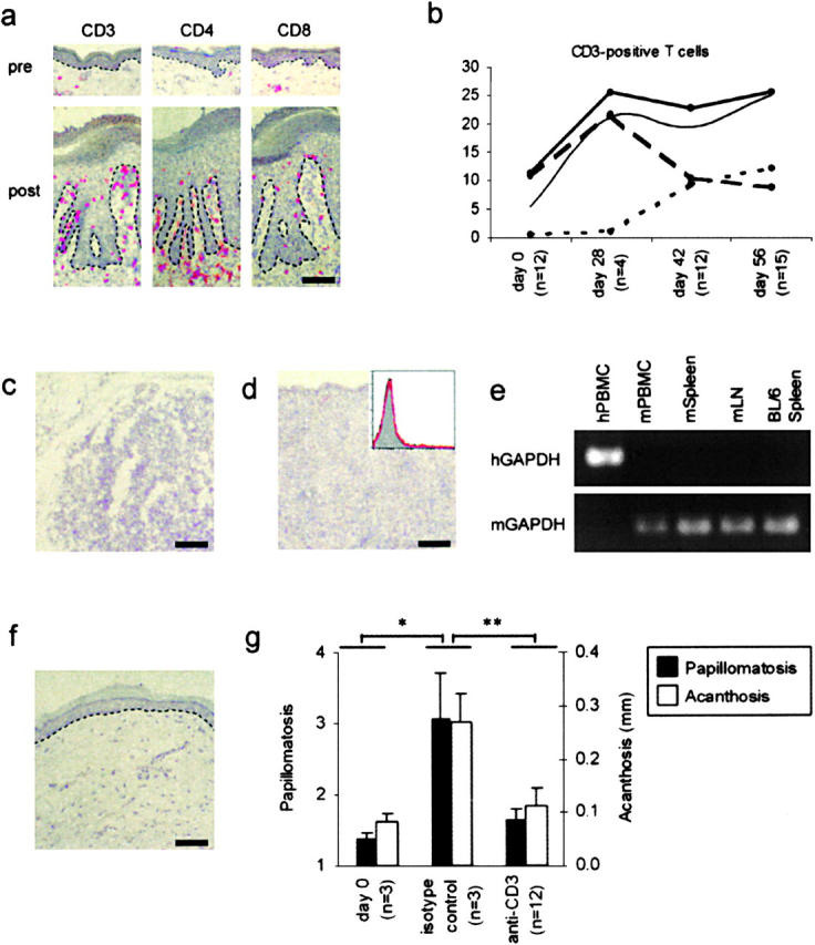
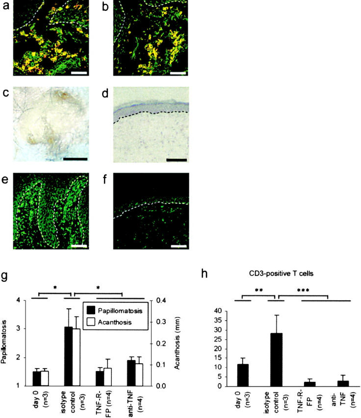
Psoriasis is a common T cell-mediated autoimmune disorder where primary onset of skin lesions is followed by chronic relapses. Progress in defining the mechanism for initiation of pathological events has been hampered by the lack of a relevant experimental model in which psoriasis develops spontaneously. We present a new animal model in which skin lesions spontaneously developed when symptomless prepsoriatic human skin was engrafted onto AGR129 mice, deficient in type I and type II interferon receptors and for the recombination activating gene 2. Upon engraftment, resident human T cells in prepsoriatic skin underwent local proliferation. T cell proliferation was crucial for development of a psoriatic phenotype because blocking of T cells led to inhibition of psoriasis development. Tumor necrosis factor-alpha was a key regulator of local T cell proliferation and subsequent disease development. Our observations highlight the importance of resident T cells in the context of lesional tumor necrosis factor-alpha production during development of a psoriatic lesion. These findings underline the importance of resident immune cells in psoriasis and will have implications for new therapeutic strategies for psoriasis and other T cell-mediated diseases.


Figures

Figure 1.
Figure 1.
Development of a psoriatic phenotype in symptomless prepsoriatic skin after transplantation onto AGR129 mice. Microscopic (A) and macroscopic (B) appearance of PN skin on the day of transplantation was comparable to normal human skin. PN skin 6–8 wk after transplantation onto AGR129 mice developed typical histological features of psoriasis such as parakeratosis (nucleated keratinocytes in stratum corneum), focal loss of the granular cell layer, acanthosis (hyperplasia of viable epidermis), papillomatosis (elongation of dermal papillae), prominent vessels of the papillary dermis, and numerous mononuclear cells representing immune cells (C). Typical clinical signs of psoriasis such as elevation, erythema, induration, and scaling could be observed (D). Histology of a biopsy of a psoriasis lesion from the same patient donating the PN skin graft (E). Control grafting experiments such as PN skin grafts onto GR129 mice (GR) (F) and skin from healthy individuals (NN skin) onto AGR129 mice (G) 8 wk after transplantation. The microscopic changes were quantified using papillomatosis and acanthosis indices of PN skin transplants onto AGR129 mice before transplantation (day 0) and on day 28, 42, and 56 after transplantation (H). Controls were skin grafts 56 d after transplantation: PN skin transplants onto RAG-2−/− mice (PN onto RAG), AR129 mice (PN onto AR), and GR129 mice (PN onto GR), and NN skin from three healthy individuals onto AGR129 mice (NN onto AGR) (H). Dashed lines indicate border between epidermis above and dermis below. n, number of grafted mice. Error bars represent one SD. Bars: (A, C, and E–G) 100 μm; (B and D) 2 mm. n.s., not significant. *, P = 0.0002; **, P = 0.005, unpaired t test.
Figure 2.
Figure 2.
Epidermal hyperproliferation, blood vessel formation, and immune activation. In contrast to the appearance of PN skin before transplantation (pre), 6–8 wk after transplantation onto AGR129 mice (post) keratin 16 was expressed throughout the epidermis and involucrin-positive cell layers were increased, indicating aberrant epidermal differentiation. Whereas before no marked expression of intercellular adhesion molecule-1 (ICAM-1) and MHC class II by keratinocytes could by observed, transplantation onto AGR129 mice and development of a psoriatic phenotype led to up-regulation of these markers, indicating activation of keratinocytes (ICAM-1, arrowhead). Strong suprabasal expression of Ki-67 demonstrated a hyperproliferative epidermal compartment in addition to proliferating dermal cells. Marked new blood vessel formation was shown by platelet endothelial cell adhesion molecule-1 (PECAM-1) staining. An activated immune compartment was observed including expanded CD1d-positive APCs in the epidermis (arrows) and dermis as well as proinflammatory cytokines such as TNF-α, IL-12, and IFN-γ. Dashed lines indicate border between epidermis above and dermis below. Bars, 50 μm.
Figure 3.
Figure 3.
Resident T cells during development of lesions. CD3, CD4, and CD8 immunostaining of PN skin before transplantation (pre) compared with 6–8 wk after transplantation onto AGR129 mice (post) (A). Epidermal (B, dotted line), dermal (B, dashed line), and total (B, thick solid line) CD3 counts in PN skin grafts before transplantation (day 0) and on day 28, 42, and 56 after transplantation onto AGR129 mice. The thin solid line indicates acanthosis index. CD3 immunostaining of LN (C), spleen (D), and flow cytometry of PBMCs (D, insert; isotype control, gray area; CD3 staining, red line) from AGR129 mice 56 d after transplantation of PN skin grafts. RT-PCR analyses of cDNA from human PBMCs (hPBMC), mouse PBMCs (mPBMC), mouse spleen (mSpleen), and mouse LNs (mLN) from AGR129 mice 56 d after transplantation of PN skin using primers for human GAPDH (hGAPDH) and mouse GAPDH (mGAPDH) (E). Spleen of untreated C57BL/6 mice (BL/6 Spleen) served as control to show specificity of the primers used. Hematoxylin staining of PN skin grafted onto AGR129 mouse and blocking of T cells with an anti–human CD3 mAb for 8 wk (F). Papillomatosis and acanthosis indices of PN skin transplants onto AGR129 mice before transplantation (day 0), 56 d after transplantation and administration of isotype-matched antibody (isotype control) or of an anti–human CD3 mAb (anti-CD3) (G). Dashed lines, n, and error bars are defined as in the legend to Fig. 1. Bars, 70 μm. *, P < 0.01; **, P < 0.0001, unpaired t test.
Figure 4.
Figure 4.
TNF-α is crucial for local T cell proliferation and psoriasis development. APCs appeared yellow in the confocal laser scanning microscope as a consequence of colocalization of specific markers CD1c (A, red) or CD83 (B, red), respectively, and TNF-α (green). Application of neutralizing anti–human TNF-α mAb or TNF receptor fusion protein over 8 wk led to the inhibition of psoriatic phenotype development (C and D). Immunostaining for TNF-α in PN skin grafts 8 wk after transplantation onto AGR129 mice and psoriasis development (E) and in PN skin grafts from mice receiving anti–TNF-α treatment (F). Papillomatosis and acanthosis indices of PN skin transplants onto AGR129 mice before transplantation (day 0) and 56 d after transplantation and administration of isotype-matched antibody (isotype control) of a TNF receptor fusion protein (TNF-R-FP) or an anti–TNF-α mAb (anti-TNF) (G). Total CD3 counts in PN skin grafts before transplantation (day 0) and 56 d after transplantation and administration of isotype-matched antibody (isotype control) of a TNF receptor fusion protein (TNF-R-FP) or an anti–TNF-α mAb (anti-TNF) (H). Dashed lines, n, and error bars are defined as in the legend to Fig. 1. Bars: (A, B, and D–F) 100 μm; (C) 2 mm. *, P = 0.01; **, P < 0.05; ***, P < 0.004, unpaired t test.

References

    1. Griffiths, C.E., and J.J. Voorhees. 1996. Psoriasis, T cells and autoimmunity. J. R. Soc. Med. 89:315–319. - PMC - PubMed
    1. Krueger, J.G. 2002. The immunologic basis for the treatment of psoriasis with new biologic agents. J. Am. Acad. Dermatol. 46:1–23. - PubMed
    1. Barker, J.N. 1991. The pathophysiology of psoriasis. Lancet. 338:227–230. - PubMed
    1. Gottlieb, S.L., P. Gilleaudeau, R. Johnson, L. Estes, T.G. Woodworth, A.B. Gottlieb, and J.G. Krueger. 1995. Response of psoriasis to a lymphocyte-selective toxin (DAB389IL-2) suggests a primary immune, but not keratinocyte, pathogenic basis. Nat. Med. 1:442–447. - PubMed
    1. Ellis, C.N., and G.G. Krueger. 2001. Treatment of chronic plaque psoriasis by selective targeting of memory effector T lymphocytes. N. Engl. J. Med. 345:248–255. - PubMed

Publication types

MeSH terms