. 2004 Mar 1;199(5):649-59.
doi: 10.1084/jem.20031097. Epub 2004 Feb 23.

Diacylated sulfoglycolipids are novel mycobacterial antigens stimulating CD1-restricted T cells during infection with Mycobacterium tuberculosis

Affiliations

Diacylated sulfoglycolipids are novel mycobacterial antigens stimulating CD1-restricted T cells during infection with Mycobacterium tuberculosis

Martine Gilleron et al. J Exp Med. .

Abstract

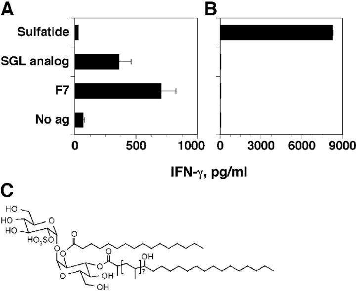
Mycobacterial lipids comprise a heterogeneous group of molecules capable of inducing T cell responses in humans. To identify novel antigenic lipids and increase our understanding of lipid-mediated immune responses, we established a panel of T cell clones with different lipid specificities. Using this approach we characterized a novel lipid antigen belonging to the group of diacylated sulfoglycolipids purified from Mycobacterium tuberculosis. The structure of this sulfoglycolipid was identified as 2-palmitoyl or 2-stearoyl-3-hydroxyphthioceranoyl-2'-sulfate-alpha-alpha'-D-trehalose (Ac2SGL). Its immunogenicity is dependent on the presence of the sulfate group and of the two fatty acids. Ac2SGL is mainly presented by CD1b molecules after internalization in a cellular compartment with low pH. Ac2SGL-specific T cells release interferon gamma, efficiently recognize M. tuberculosis-infected cells, and kill intracellular bacteria. The presence of Ac2SGL-responsive T cells in vivo is strictly dependent on previous contact with M. tuberculosis, but independent from the development of clinically overt disease. These properties identify Ac2SGL as a promising candidate to be tested in novel vaccines against tuberculosis.


Figures

Figure 1.
Figure 1.
Characterization of the T cell antigen. Negative MALDI-Tof mass spectra of (A) the “acetone-soluble” fraction (fraction 1) and (B) fraction 7 obtained after silicic acid column chromatography containing the stimulatory activity. See Table S1 for peak interpretation. Inset: Structural model of the M. tuberculosis SGL with R corresponding to hydroxyphthioceranoic, palmitic, or stearic acid. Ac4SGL contains three hydroxyphthioceranoic acids and one palmitic or stearic acid, Ac3SGL contains two hydroxyphthioceranoic acids and one palmitic or stearic acid, and Ac2SGL contains one hydroxyphthioceranoic acid and one palmitic or stearic acid. MS peaks are labeled with mass values calculated using C = 12, H = 1, O = 16, P = 31, and S = 32.
Figure 2.
Figure 2.
Identification of the active ligand present in fraction 7 as Ac2SGL. (A) TLC analysis of the components present in fraction 7 after reverse chromatography purification revealed by orcinol. Numbers indicate the subfractions tested in the following panels. (B) Negative MALDI-Tof mass spectra of the subfractions 7.1, 7.3, 7.4, and 7.5. See Table S1 for peak interpretation. (C) Stimulation of the Z4B27 T cell clone by (•) subfraction 7.1 from A, (▵) subfraction 7.3, (⋄) subfraction 7.4, (□) subfraction 7.5, (▪) desulfatated Ac2SGL, and (○) control active fraction 7 as measured by IFN-γ ELISA. Bars indicate SD.
Figure 3.
Figure 3.
Structure determination of Ac2SGL by 1D 1H NMR spectrum analysis (δ 1H: 0–7.8). The structure of Ac2SGL is shown in the inset. The two glucose units are labeled I and II, as indicated in the inset. R corresponds to hydroxyphthioceranoic, palmitic, or stearic acid.
Figure 4.
Figure 4.
Biological activity of a synthetic analogue and structure of the natural active molecule. (A) Activation of the Ac2SGL-specific T cell clone Z4B27 using synthetic 2,3-dipalmitoyl-α-α'-d-trehalose-2′-sulfate (SGL analogue). Purified Ac2SGL and synthetic Ac2SGL analogue were used at 10 μg/ml. Release of IFN-γ was tested by ELISA. The results are representative of two experiments. Bars indicate SD. (B) Control with sulfatide-specific DS1C9b T cell clone. Sulfatide was used at 10 μg/ml. Bars indicate SD. (C) Structure of M. tuberculosis Ac2SGL. The presented structure corresponds to one of the major Ac2SGL acyl forms (mol wt 1,250).
Figure 5.
Figure 5.
CD1b-restricted presentation of Ac2SGL. (A) Autologous CD1+ APCs and not PBMCs or EBV-transformed lymphoblastoid cell lines present Ac2SGL. (B) Presentation by CD1+ APCs isolated from different individuals and by cell lines of the monocytic lineage. (C) Inhibition of T cell activation by anti-CD1b mAbs. (D) Presentation by CD1b-transfected THP-1 cells. Release of IFN-γ by the Z4B27 T cell clone was measured by ELISA. White bars represent response in the absence of antigen. A second Ac2SGL-specific and CD1b-restricted T cell clone gave similar results (unpublished data). Bars indicate SD.
Figure 6.
Figure 6.
Requirements for presentation of Ac2SGL. (A) Presentation of Ac2SGL is inhibited when APCs are pulsed with Ac2SGL (solid bars) at 4°C, or in the presence of monensin or chloroquine. White bars represent response in the absence of antigen. (C) APCs pulsed and then fixed present Ac2SGL. (B and D) The same treatments did not affect presentation of sulfatide to the CD1b-restricted and sulfatide-specific DS1C9b T cell clone. (A and C) Release of IFN-γ by the Z4B27 T cell clone measured by ELISA. Similar results were obtained in three additional experiments and also with the Z4A26 T cell clone (unpublished data). Medium indicates response in the absence of treatment. Bars indicate SD.
Figure 7.
Figure 7.
Ac2SGL-specific T cells recognize and kill M. tuberculosis H37Rv-infected cells. CD1+ APCs were pulsed with Ac2SGL or infected with virulent M. tuberculosis and used to stimulate the Z4B27 (A) or the Z4A26 (B) T cell clones. Released IFN-γ was detected by ELISA. The results are representative of four independent experiments. Bars indicate SD. (C) Ac2SGL-specific T cells kill intracellular M. tuberculosis. CD1+ APCs were infected with virulent M. tuberculosis. Z4B27 T cells were added at different ratios as indicated. After 72 h of incubation, living intracellular bacteria were counted as described in Materials and Methods. Average CFU ± SEM calculated from four dilutions, each performed in duplicate are shown. Similar results were obtained in two additional experiments.
Figure 8.
Figure 8.
Response of PBMCs to Ac2SGL ex vivo and inhibition with anti-CD1 and anti-MHC mAbs. (A) PBMCs from healthy donors (PPD+ or PPD) and from patients with pulmonary tuberculosis were cultured in the presence of autologous CD1+ APCs overnight pulsed with Ac2SGL. Supernatants were harvested and IFN-γ content was measured by ELISA. Each dot presents an individual donor and the dashed line depicts the detection threshold of 15 pg/ml of this assay. Unstimulated samples served as controls and IFN-γ was regularly below the level of detection. (B) This graph summarizes the results ± SEM from three PPD+ healthy donors using fraction 7 (10 μg/ml) as antigen. IFN-γ production in the individual experiments was 473, 357, and 349 pg/ml.

References

    1. Kaufmann, S.H. 2001. How can immunology contribute to the control of tuberculosis? Nat. Rev. Immunol. 1:20–30. - PubMed
    1. Gumperz, J.E., and M.B. Brenner. 2001. CD1-specific T cells in microbial immunity. Curr. Opin. Immunol. 13:471–478. - PubMed
    1. Vincent, M.S., J.E. Gumperz, and M.B. Brenner. 2003. Understanding the function of CD1-restricted T cells. Nat. Immunol. 4:517–523. - PubMed
    1. Flynn, J.L., and J. Chan. 2001. Immunology of tuberculosis. Annu. Rev. Immunol. 19:93–129. - PubMed
    1. Stenger, S., and R.L. Modlin. 1999. T cell mediated immunity to Mycobacterium tuberculosis. Curr. Opin. Microbiol. 2:89–93. - PubMed

Publication types