. 2004 Mar 2;101(9):2858-63.
doi: 10.1073/pnas.0400044101. Epub 2004 Feb 23.

Determinants of liposome fusion mediated by synaptic SNARE proteins

Affiliations

Determinants of liposome fusion mediated by synaptic SNARE proteins

Christina G Schuette et al. Proc Natl Acad Sci U S A. .

Abstract

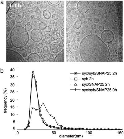
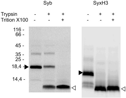
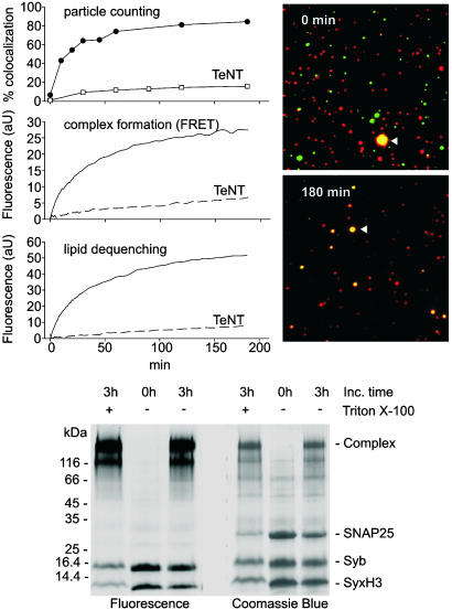
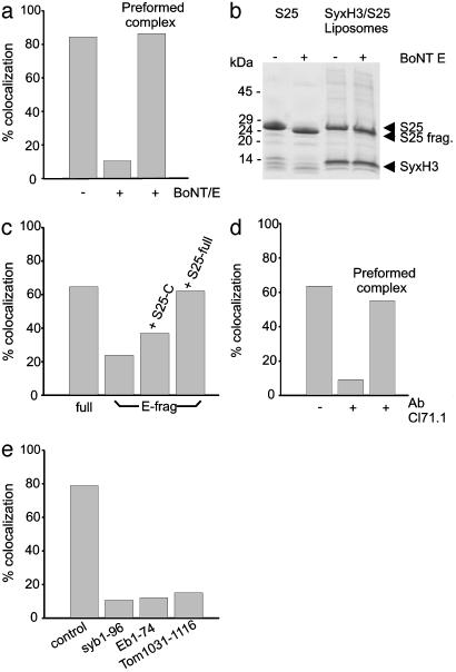
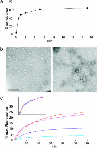
Synaptic exocytosis requires the soluble N-ethylmaleimide-sensitive factor attachment protein receptor (SNARE) proteins syntaxin 1, SNAP-25, and synaptobrevin (VAMP). Assembly of the SNAREs into a stable core complex is supposed to catalyze membrane fusion, and proteoliposomes reconstituted with synaptic SNARE proteins spontaneously fuse with each other. We now show that liposome fusion mediated by synaptic SNAREs is inhibited by botulinum neurotoxin E (BoNT/E) but can be rescued by supplementing the C-terminal portion of SNAP-25. Furthermore, fusion is prevented by a SNAP-25-specific antibody known to block exocytosis in chromaffin cells, and it is competed for by soluble fragments of the R-SNAREs synaptobrevin 2, endobrevin/VAMP-8, and tomosyn. No accumulation of clustered vesicles is observed during the reaction. Rapid artificial clustering of SNARE-containing proteoliposomes enhances the fusion rate at low but not at saturating liposome concentrations. We conclude that the rate of liposome fusion is dominated by the intrinsic properties of the SNAREs rather than by the preceding docking step.


Figures

Fig. 3.
Fig. 3.
Increase of liposome size after fusion, analyzed by cryo-electron microscopy. (a) Images of a fusion reaction shock-frozen either immediately (t = 0 h) or 2 h (t = 2 h) after the begin of the reaction. (Bar = 100 nm.) (b) Distribution of the liposome diameters (n ≈ 1,000) at the beginning (0 h) and end (2 h) of the reaction, in comparison with the diameter distribution of separately incubated donor and acceptor liposomes.
Fig. 1.
Fig. 1.
Orientation of the SNAREs in proteoliposomes. Liposomes reconstituted with either synaptobrevin labeled with Alexa594 (syb) or a preformed complex of syntaxin H3 (labeled with Alexa488, SyxH3) and SNAP-25 were incubated with trypsin in absence or presence of Triton X-100 and analyzed by SDS/PAGE and fluorescence imaging. Filled and open arrowheads mark the position of undigested protein and protease-resistant fragments, respectively.
Fig. 2.
Fig. 2.
(Upper Left) Comparison of fusion kinetics using particle counting (Top), FRET (Middle), and lipid dequenching (Bottom). Donor and acceptor liposomes were coreconstituted with syntaxin H3/SNAP-25 and synaptobrevin, respectively. For particle counting and FRET, C-terminally labeled variants of syntaxin-H3 and synaptobrevin were used. Fusion was performed at 37°C. As control, synaptobrevin liposomes were preincubated for 30 min with 3 μM tetanus toxin light chain (TeNT) at 37°C. (Upper Right) Representative microscopic pictures of a fusion assay at the begin (Upper) and end (Lower) of the incubation. The pictures acquired in the red and green channel were aligned by using TetraSpeck beads (arrowheads) as reference. (Bottom) Assembly status of SNAREs at the beginning (0 h) and end (3 h) of the fusion reaction using fluorescently labeled syntaxin and synaptobrevin. As control, one sample was incubated for 3 h at 37°C in the presence of 2% (vol/vol) of Triton X-100 to allow for maximal complex formation. The samples were separated by SDS/PAGE and visualized either by fluorescence imaging using a filter set consisting of HQ470/40 excitation filter and a HQ645/75 emission filter (Left) or by Coomassie staining (Right).
Fig. 4.
Fig. 4.
Perturbation of SNARE assembly leads to a reduction of fusion. All assays were analyzed by particle counting. (a) Effect of BoNT/E on liposome fusion. Toxin treatment was carried out either with isolated SNAP-25 that was then added to syntaxin-containing liposomes, or with liposomes coreconstituted with preformed syxH3/SNAP-25 complexes. All preincubations were for 30 min at 37°C, and fusion was analyzed after 3 h. (b) SNAP-25 is resistant to BoNT/E cleavage in preformed syxH3/SNAP-25 complexes. Liposomes containing preformed complexes were incubated with BoNT/E light chain in a 1:50 molar ratio of toxin to SNAP-25 for 1 h at 37°C. As control, free SNAP-25 was incubated in parallel. Samples before and after treatment were separated by SDS/PAGE and visualized by Coomassie blue staining. S25, SNAP-25, S25 frag., SNAP-25 fragment caused by BoNT/E cleavage. (c) Both intact SNAP-25 (S25-full) and the C-terminal SNARE motif of SNAP-25 (S25-C) rescue fusion of liposomes containing C-terminally truncated SNAP-25 [BoNT/E fragment (E-frag), amino acid 1–180]. Liposomes containing syxH3 were preincubated for 30 min with E-frag (control: full-length SNAP-25, Left) and then either directly used for the fusion assay or incubated for additional 30 min in the presence of S25-full or S25-C before fusion. (d) Effect of monoclonal antibody Cl 71.1 (directed against the N-terminal SNARE-motif of SNAP-25) on fusion. Liposomes containing syxH3 were incubated with SNAP-25 either in the presence of absence of antibody 71.1 for 30 min at 37°C. In parallel, liposomes containing a preformed complex of syxH3 and SNAP-25 were incubated with antibody 71.1 for 30 min at 37°C. Fusion was analyzed after 2 h. (e) Soluble R-SNARE motifs inhibit liposome fusion. To liposomes containing a preformed complex of syxH3 and SNAP-25 the soluble SNARE-motifs of synaptobrevin, endobrevin, and tomosyn were added in a 2-fold molar excess over syntaxin, and the mixture was incubated at 37°C for 30 min before starting the fusion reaction. Fusion was analyzed after 3 h.
Fig. 5.
Fig. 5.
Influence of preclustering on the liposome fusion rate. (a) Determination of the clustering rate by particle counting. Streptavidin-saturated and -free biotinylated liposomes containing synaptobrevin labeled with Alexa594 and Alexa488, respectively, were mixed, and the reaction was terminated by 1:10 dilution in 50 mM biotin. (b) Electron microscopy of negatively stained vesicles after artificial docking (Right). For comparison, streptavidin saturated liposomes adjusted to the same liposome concentration are shown (Left). (Bar = 200 nm.) (c) Effect of docking on fusion kinetics under standard conditions and at 100-fold dilution of liposomes. Donor liposomes containing syxH3/SNAP-25 were either saturated with streptavidin or streptavidin was omitted so that docking could not occur. Acceptor liposomes contained synaptobrevin and free biotin-phycoerythrin. Red, standard conditions with streptavidin; pink, standard conditions without streptavidin; dark blue, 100-fold dilution with streptavidin; light blue, 100-fold dilution without streptavidin. Fusion was monitored by lipid dequenching. (Inset) Normalized fusion rates of predocked vesicles show almost identical fusion kinetics.

References

    1. Chen, Y. A. & Scheller, R. H. (2001) Nat. Rev. Mol. Cell. Biol. 2, 98-106. - PubMed
    1. Jahn, R., Lang, T. & Sudhof, T. C. (2003) Cell 112, 519-533. - PubMed
    1. Rizo, J. & Sudhof, T. C. (2002) Nat. Rev. Neurosci. 3, 641-653. - PubMed
    1. Fasshauer, D., Sutton, R. B., Brunger, A. T. & Jahn, R. (1998) J. Biol. Chem. 95, 15781-15786. - PMC - PubMed
    1. Sutton, B., Fasshauer, D., Jahn, R. & Brünger, A. T. (1998) Nature 395, 347-353. - PubMed

Publication types

LinkOut - more resources