. 2004 Mar 2;101(9):2817-22.
doi: 10.1073/pnas.0400062101. Epub 2004 Feb 23.

Tissue damage in the amyloidoses: Transthyretin monomers and nonnative oligomers are the major cytotoxic species in tissue culture

Affiliations

Tissue damage in the amyloidoses: Transthyretin monomers and nonnative oligomers are the major cytotoxic species in tissue culture

Natàlia Reixach et al. Proc Natl Acad Sci U S A. .

Abstract

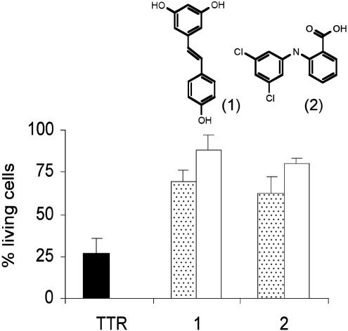
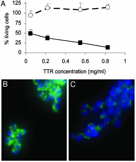
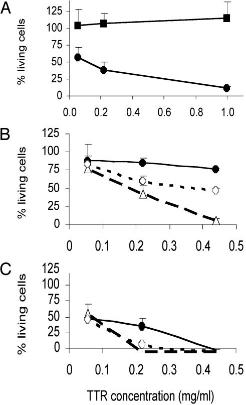
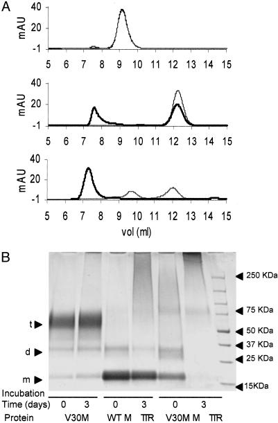
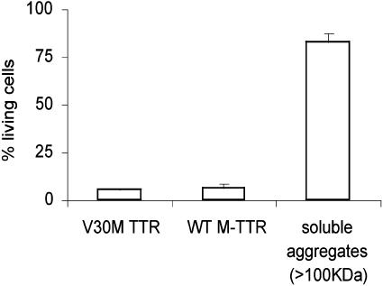
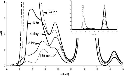
The transthyretin (TTR) amyloidoses are human diseases in which the misfolded TTR protein aggregates in tissues with subsequent visceral, peripheral, and autonomic nerve dysfunction. Recent reports have stressed the importance of oligomeric intermediates as major cytotoxic species in various forms of amyloidogenesis. We have examined the cytotoxic effects of several quaternary structural states of wild-type and variant TTR proteins on cells of neural lineage. TTR amyloid fibrils and soluble aggregates >100 kDa were not toxic. Incubation of TTR under the conditions of the cell assay and analysis by size-exclusion chromatography and SDS/PAGE reveal that monomeric TTR or relatively small, rapidly formed aggregates of a maximum size of six subunits were the major cytotoxic species. Small molecules that stabilize the native tetrameric state were shown to prevent toxicity. The studies are consistent with a model in which the misfolded TTR monomer rapidly aggregates to form transient low molecular mass assemblies (<100 kDa) that are highly cytotoxic in tissue culture.


Figures

Fig. 2.
Fig. 2.
V30M TTR-induced cytotoxicity can be inhibited by compounds that stabilize TTR tetramer. Cells treated with V30M TTR (black bar), an equimolar mixture of V30M TTR and compounds (dotted bars), and compounds alone (white bars). Bars represent the SD of triplicate determinations.
Fig. 1.
Fig. 1.
(A) Cytotoxicity of WT TTR (○) and V30M TTR (▪) on IMR-32 cells after 3 days of incubation at 37°C. Bars represent the SD of triplicate determinations. (B and C) Fluorescent microscopy images of IMR-32 cells stained with Rhodamine 123 (green) and Hoechst 33342 (blue) after 40 h incubation with 1 mg/ml V30M TTR. (B) Control cells (Opti-MEM only). (C) V30M TTR-treated cells. The decrease in mitochondrial membrane potential (green) of TTR-treated cells compared with control cells, suggest that the TTR triggers apoptosis in IMR-32 cells. Some nuclei condensation (clear blue) is seen also after TTR treatment.
Fig. 3.
Fig. 3.
Cytotoxicity of quaternary structure variants of TTR in IMR-32. (A) V30M TTR fibrils (▪) versus soluble V30M TTR (•), incubated for 3 days. (B and C) V30M TTR (•) versus WT M-TTR (○) and V30M M-TTR (○), incubated for 36 h (B) or 3 days (C) at 37°C. Bars represent the SD of triplicate determinations.
Fig. 4.
Fig. 4.
(A) SEC analysis (10/30 Superdex 75 column) of V30M TTR (Top), WT M-TTR (Middle), and V30M M-TTR (Bottom) at 0.44 mg/ml incubated at 37°C in Opti-MEM. Gray line, time 0; black line, analysis of protein solutions after 3 days of incubation. At Top (V30M TTR analysis), the two curves (time 0 and time 3 days) are superimposed. (B) SDS/PAGE of the same protein solutions than in A after cross-linking with glutaraldehyde. From left to right, V30M TTR (0 and 3 days), WT M-TTR (0 and 3 days), and V30M M-TTR (0 and 3 days) are shown. Arrowheads at right indicate the molecular weight standards. Arrowheads at left indicate sizes of monomer (m), dimer (d), and tetramer (t).
Fig. 5.
Fig. 5.
Cytotoxicity of 1-mg/ml solutions of V30M TTR, WT M-TTR, and soluble aggregates (>100 kDa) derived from WT M-TTR in IMR-32 cells after 3 days of incubation at 37°C.
Fig. 6.
Fig. 6.
SEC analysis (10/30 Superdex 75 column) of the aggregation process of WT M-TTR at 1 mg/ml in Opti-MEM at 37°C show small aggregates forming (10.2, 9.8, and 8.8 ml). (Inset) Full chromatograms. The species seen at 10.2 ml disappears after1hof incubation. The species seen at 9.8 ml reaches a maximum between 6 and 12 h, and decreases afterward. The species at 8.8 ml increases with time.

References

    1. Buxbaum, J. N. & Tagoe, C. E. (2000) Annu. Rev. Med. 51, 543-569. - PubMed
    1. Merlini, G. & Bellotti, V. (2003) N. Engl. J. Med. 349, 583-596. - PubMed
    1. Sacchettini, J. C. & Kelly, J. W. (2002) Nat. Rev. Drug Discov. 1, 267-275. - PubMed
    1. Kelly, J. W. (1998) Curr. Opin. Struct. Biol. 8, 101-106. - PubMed
    1. Damas, A. M. & Saraiva, M. J. (2000) J. Struct. Biol. 130, 290-299. - PubMed

Publication types