Identification of a target RNA motif for RNA-binding protein HuR
- PMID: 14981256
- PMCID: PMC365732
- DOI: 10.1073/pnas.0306453101
Identification of a target RNA motif for RNA-binding protein HuR
Abstract
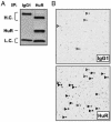
HuR, a protein that binds to specific mRNA subsets, is increasingly recognized as a pivotal posttranscriptional regulator of gene expression. Here, HuR was immunoprecipitated under conditions that preserved HuR-RNA interactions, and HuR-bound target mRNAs were identified by cDNA array hybridization. Analysis of primary sequences and secondary structures shared among HuR targets led to the identification of a 17- to 20-base-long RNA motif rich in uracils. This HuR motif was found in almost all mRNAs previously reported to be HuR targets, was located preferentially within 3' untranslated regions of all unigene transcripts examined, and was conserved in >50% of human and mouse homologous genes. Importantly, the HuR motif allowed the successful prediction and subsequent validation of novel HuR targets from gene databases. This study describes an HuR target RNA motif and presents a general strategy for identifying target motifs for RNA-binding proteins.
Figures


Similar articles
-
Global analysis of HuR-regulated gene expression in colon cancer systems of reducing complexity.Gene Expr. 2004;12(1):49-59. doi: 10.3727/000000004783992215. Gene Expr. 2004. PMID: 15473260 Free PMC article.
-
Elucidation of a C-rich signature motif in target mRNAs of RNA-binding protein TIAR.Mol Cell Biol. 2007 Oct;27(19):6806-17. doi: 10.1128/MCB.01036-07. Epub 2007 Aug 6. Mol Cell Biol. 2007. PMID: 17682065 Free PMC article.
-
The RNA binding protein HuR differentially regulates unique subsets of mRNAs in estrogen receptor negative and estrogen receptor positive breast cancer.BMC Cancer. 2010 Apr 6;10:126. doi: 10.1186/1471-2407-10-126. BMC Cancer. 2010. PMID: 20370918 Free PMC article.
-
HuR and mRNA stability.Cell Mol Life Sci. 2001 Feb;58(2):266-77. doi: 10.1007/PL00000854. Cell Mol Life Sci. 2001. PMID: 11289308 Free PMC article. Review.
-
Mechanisms coordinating ELAV/Hu mRNA regulons.Curr Opin Genet Dev. 2013 Feb;23(1):35-43. doi: 10.1016/j.gde.2012.12.006. Epub 2013 Jan 9. Curr Opin Genet Dev. 2013. PMID: 23312841 Free PMC article. Review.
Cited by
-
The RNA binding protein HuR determines the differential translation of autism-associated FoxP subfamily members in the developing neocortex.Sci Rep. 2016 Jul 7;6:28998. doi: 10.1038/srep28998. Sci Rep. 2016. PMID: 27383233 Free PMC article.
-
An endoplasmic reticulum stress-regulated lncRNA hosting a microRNA megacluster induces early features of diabetic nephropathy.Nat Commun. 2016 Sep 30;7:12864. doi: 10.1038/ncomms12864. Nat Commun. 2016. PMID: 27686049 Free PMC article.
-
HuR inhibits apoptosis by amplifying Akt signaling through a positive feedback loop.J Cell Physiol. 2013 Jan;228(1):182-9. doi: 10.1002/jcp.24120. J Cell Physiol. 2013. PMID: 22674407 Free PMC article.
-
Bioinformatic identification of novel elements potentially involved in messenger RNA fate control during spermatogenesis.Biol Reprod. 2012 Dec 13;87(6):138. doi: 10.1095/biolreprod.112.102434. Print 2012 Jun. Biol Reprod. 2012. PMID: 23053435 Free PMC article.
-
Enhanced proliferation of cultured human vascular smooth muscle cells linked to increased function of RNA-binding protein HuR.J Biol Chem. 2005 Jun 17;280(24):22819-26. doi: 10.1074/jbc.M501106200. Epub 2005 Apr 11. J Biol Chem. 2005. PMID: 15824116 Free PMC article.
References
MeSH terms
Substances
LinkOut - more resources
Full Text Sources
Other Literature Sources
Molecular Biology Databases
Miscellaneous

