. 2004 Mar 2;101(9):2712-7.
doi: 10.1073/pnas.0308612100. Epub 2004 Feb 23.

The solution structure of the N-terminal domain of E3L shows a tyrosine conformation that may explain its reduced affinity to Z-DNA in vitro

Affiliations

The solution structure of the N-terminal domain of E3L shows a tyrosine conformation that may explain its reduced affinity to Z-DNA in vitro

Jan D Kahmann et al. Proc Natl Acad Sci U S A. .

Abstract

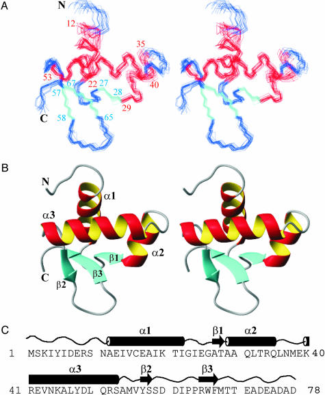
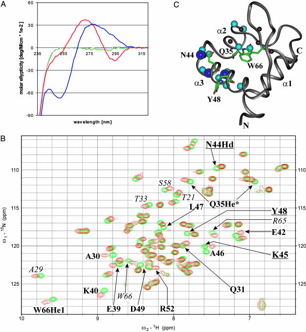
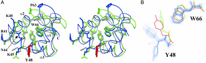
The N-terminal domain of the vaccinia virus protein E3L (Z alpha(E3L)) is essential for full viral pathogenicity in mice. It has sequence similarity to the high-affinity human Z-DNA-binding domains Z alpha(ADAR1) and Z alpha(DLM1). Here, we report the solution structure of Z alpha(E3L) and the chemical shift map of its interaction surface with Z-DNA. The global structure and the Z-DNA interaction surface of Z alpha(E3L) are very similar to the high-affinity Z-DNA-binding domains Z alpha(ADAR1) and Z alpha(DLM1). However, the key Z-DNA contacting residue Y48 of Z alpha(E3L) adopts a different side chain conformation in unbound Z alpha(E3L), which requires rearrangement for binding to Z-DNA. This difference suggests a molecular basis for the significantly lower in vitro affinity of Z alpha(E3L) to Z-DNA compared with its homologues.


Figures

Fig. 1.
Fig. 1.
3D solution structure of ZαE3L.(A) Stereoview of the ensemble of the 20 lowest energy structures of E3L. The first and last residue of the 3 α-helices (red) (α1, α2, α3) and 3 β-strands (cyan) (β1, β2, β3) are numbered. (B) Stereoview of the backbone ribbon of the mean structure illustrating the (α plus β) helix–turn–helix fold of ZαE3L. The N- and C-termini are labeled with N and C, respectively. (C) The secondary structure of ZαE3L is shown with the amino acid sequence directly under it.
Fig. 2.
Fig. 2.
(A) The conformation of substrate DNA in the presence of ZαE3L. The CD spectrum of d(CG)6T4(CG)6 substrate DNA in the presence of ZαE3L (blue curve) shows a conventional B-DNA conformation. In contrast, the CD spectrum of d(CG)6T4(CG)6 in the presence of ZαE3L and [Co(NH3)6]3+, which is known to promote the conversion from B- to Z-DNA (17), shows an inversion of ellipticity at 295 and 255 nm characteristic for duplex DNA in the Z-conformation (red curve). For control, ZαE3L alone (green curve) and ZαE3L in the presence of [Co(NH3)6]3+ (gray curve) show zero ellipticity between 320 and 250 nm. (B) Chemical shift map of the ZαE3L/substrate DNA interaction. The 15N-HSQC NMR spectrum of ZαE3L in the presence of d(CG)6T4(CG)6 and [Co(NH3)6]3+ (red peaks) shows several large chemical shift changes compared with ZαE3L alone (green peaks). In addition, there are three vanishing resonances (residues underlined). This finding indicates a selective interaction between ZαE3L and d(CG)6T4(CG)6 in the vicinity of the affected residues. The 15N-HSQC NMR spectrum of ZαE3L in the presence of d(CG)6T4(CG)6 substrate DNA shows no chemical shift alterations as compared with ZαE3L alone (Fig. 4), indicating no discernible interaction under these conditions. Further, the spectrum of ZαE3L in the presence of [Co(NH3)6]3+ is identical to ZαE3L alone (not shown). (C) The Z-DNA-binding site of ZαE3L. In the presence of d(CG)6T4(CG)6 and [Co(NH3)6]3+, the H atoms of ZαE3L that show large averaged chemical shift changes (≥0.082 ppm; bold labels in B) are shown as cyan spheres. H atoms with medium shifts (≥0.068 ppm; italic labels in B) are shown as dark red spheres. Atoms of ZαE3L that show vanishing resonances (underlined labels in B) are shown as dark blue spheres in the 3D structure of ZαE3L. Chemical shifts are listed in Table 2, which is published as supporting information on the PNAS web site. These data indicate a contiguous Z-DNA-binding site made up of helices α3 and α2 and the area around W66.
Fig. 3.
Fig. 3.
Superposition of the 3D structures of ZαE3L (blue), ZαADAR1 (light green), and ZαDLM1 (gray) in stereo. (A) The secondary structure elements of ZαE3L, ZαADAR1, and ZαDLM1 superimpose well except for the loop containing P63 (side chain labeled). Also, the side chains of K40, R41, N44, K45, and W66, which contact the bound Z-DNA in the co-crystal structures of ZαADAR1 and ZαDLM1, show very similar positions. Only the side chain of Y48 adopts a distinct position in ZαE3L (red) as compared with ZαADAR1 and ZαDLM1.(B) Distinct Y48-W66 distance between ZαE3L and ZαADAR1. Superposition of Y48 and W66 between the 20 lowest energy structures of E3L (blue), unbound ZαADAR1 (red), and bound ZαADAR1 (light green). Whereas Y48 residues are within van-der-Waals distance to W66 in both ZαADAR1 structures, it adopts two solvent exposed rotamer positions in ZαE3L, which are 7.2 and 10.8 Å apart from W66.

References

    1. Cohen, J. (2001) Science 294, 985. - PubMed
    1. Brandt, T. A. & Jacobs, B. L. (2001) J. Virol. 75, 850-856. - PMC - PubMed
    1. Kim, Y. G., Lowenhaupt, K., Oh, D. B., Kim, K. K. & Rich, A. (2004) Proc. Natl. Acad. Sci. USA, in press.
    1. Kim, Y. G., Muralinath, M., Brandt, T., Pearcy, M., Hauns, K., Lowenhaupt, K., Jacobs, B. L. & Rich, A. (2003) Proc. Natl. Acad. Sci. USA 100, 6974-6979. - PMC - PubMed
    1. Schade, M., Turner, C. J., Lowenhaupt, K., Rich, A. & Herbert, A. (1999) EMBO J. 18, 470-479. - PMC - PubMed

Publication types

Associated data

LinkOut - more resources