. 2004 Mar;164(3):817-30.
doi: 10.1016/S0002-9440(10)63170-7.

Human pancreatic islet-derived progenitor cell engraftment in immunocompetent mice

Affiliations

Human pancreatic islet-derived progenitor cell engraftment in immunocompetent mice

Elizabeth J Abraham et al. Am J Pathol. 2004 Mar.

Abstract

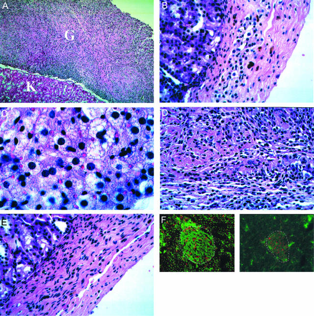
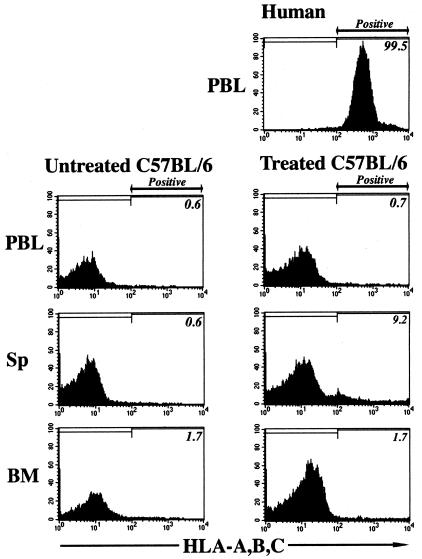
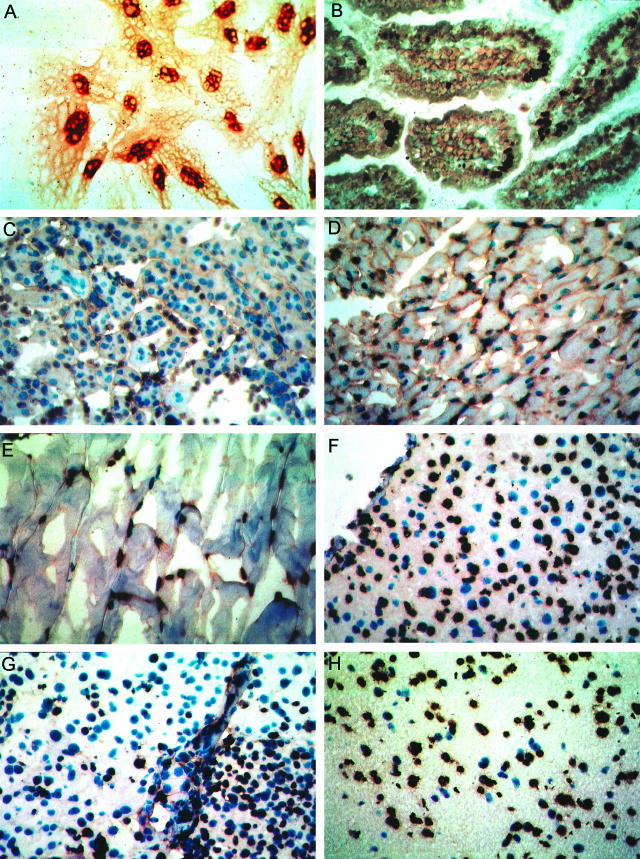
The potential for the use of stem/progenitor cells for the restoration of injured or diseased tissues has garnered much interest recently, establishing a new field of research called regenerative medicine. Attention has been focused on embryonic stem cells derived from human fetal tissues. However, the use of human fetal tissue for research and transplantation is controversial. An alternative is the isolation and utilization of multipotent stem/progenitor cells derived from adult donor tissues. We have previously reported on the isolation, propagation, and partial characterization of a population of stem/progenitor cells isolated from the pancreatic islets of Langerhans of adult human donor pancreata. Here we show that these human adult tissue-derived cells, nestin-positive islet-derived stem/progenitor cells, prepared from human adult pancreata survive engraftment and produce tissue chimerism when transplanted into immunocompetent mice either under the kidney capsule or by systemic injection. These xenografts seem to induce immune tolerance by establishing a mixed chimerism in the mice. We propose that a population of stem/progenitor cells isolated from the islets of the pancreas can cross xenogeneic transplantation immune barriers, induce tissue tolerance, and grow.


Figures

Figure 1
Figure 1
Karyotype of human NIPs. The NIPs were passaged in continuous culture for 5 months. The NIP culture was derived from pancreatic islets obtained from a male donor pancreas. The karyotype is that of a diploid (euploid) 46, XY male.
Figure 2
Figure 2
Engraftment of human NIPs in immunocompetent mice. A: Mouse kidney removed 35 days after transplantation of human NIPs under the kidney capsule. The mouse was a female C57BL/6 without immunosuppression. The whitish mass of tissue at the top of the kidney (arrow) is the expanded NIP graft, contrasting with the kidney tissue. The engraftment of NIPs has been achieved in 10 of 26 of nonimmunosuppressed mice so transplanted and examined from 15 to 60 days after the xenograft. B: A human NIP transfected with a plasmid expressing EGFP viewed under UV light. The cells expressing EGFP were transplanted under the kidney capsule of an immunocompetent mouse without immunosuppression. C: Gross morphology of the human NIP graft expressing EGFP in a kidney removed 15 days after transplantation of the NIPs. The graft (G) and kidney (K) were viewed under UV light. D: EGFP-positive cells present in explant cultures prepared from the graft shown in C (5 days of culture).
Figure 3
Figure 3
Histomorphology of a human NIP graft in an immunocompetent mouse. A: Tissue sections (H&E stained) of a C57BL/6 nonimmunosuppressed mouse kidney and human NIP graft 35 days after transplantation of human NIPs under the kidney capsule. K, Mouse kidney; G, human NIP graft. Note that the transplanted human NIP cells grow and are not rejected by the mouse recipient of the graft. B: The graft contains localized regions of glandular-like tissue (liver). C: Magnification of glandular-like liver tissue in graft. D: Mesenchymal-stromal-like tissue in the graft. E: Magnification of the stromal-like tissue in the graft. F: Nestin-positive cells in the pancreas do not express either class I or class II MHC antigens. Dual immunostaining (green) of a rat pancreas with antisera to MHC class I antigen (left) and MHC class II antigen (right). Nestin-positive cells are immunostained in red. The absence of yellow cells, which would indicate that a cell co-expresses nestin and MHC antigens, shows that NIPs do not express either MHC class I or class II antigens. Dashed lines denote boundaries of pancreatic islets. Original magnifications: ×100 (A); ×1000 (C); ×400 (E).
Figure 4
Figure 4
Immunohistochemical analyses of human NIP grafts in immunocompetent mice. A: Immunostaining for human-specific LCA, CD45, in a section of mouse kidney transplanted (kidney capsule) with human NIPs examined 35 days after the transplant. The brown-colored cells (shown in the figure as grayish-black) are the cells in the xenograft that express human LCA in the recipient mouse. K, Kidney; G, graft. B: LCA immunostaining of the graft shown at higher magnification. C: Keratin immunostaining. D: Vimentin immunostaining. E: Detection of pancreatic endocrine hormones insulin and glucagon, and pancreas- and duodenal-specific homeobox transcription factor, PDX-1 in serial sections of expanded grafts of human NIPs 15 days after transplantation under the kidney capsules of immunocompetent C57BL/6 mice. Islands of pancreatic endocrine-like tissue are found scattered about in the graft, which grew to ∼50% the volume of the kidney without evidence of invasion or oncogenicity. The top row shows two representative fields of the grafts. GP-IgG and normal rabbit serum are the control antisera for insulin and glucagon/PDX-1, respectively (bottom row). Immunostaining done by glucose oxidase method (shown in the figure as grayish-black). Immunocytochemistry was also done by diaminobenzidine (Vectastain ABC) with similar results. Asterisk denotes a burnout defect artifact in the CCD camera. Original magnifications: ×100 (A, E); ×400 (B–D).
Figure 4
Figure 4
Immunohistochemical analyses of human NIP grafts in immunocompetent mice. A: Immunostaining for human-specific LCA, CD45, in a section of mouse kidney transplanted (kidney capsule) with human NIPs examined 35 days after the transplant. The brown-colored cells (shown in the figure as grayish-black) are the cells in the xenograft that express human LCA in the recipient mouse. K, Kidney; G, graft. B: LCA immunostaining of the graft shown at higher magnification. C: Keratin immunostaining. D: Vimentin immunostaining. E: Detection of pancreatic endocrine hormones insulin and glucagon, and pancreas- and duodenal-specific homeobox transcription factor, PDX-1 in serial sections of expanded grafts of human NIPs 15 days after transplantation under the kidney capsules of immunocompetent C57BL/6 mice. Islands of pancreatic endocrine-like tissue are found scattered about in the graft, which grew to ∼50% the volume of the kidney without evidence of invasion or oncogenicity. The top row shows two representative fields of the grafts. GP-IgG and normal rabbit serum are the control antisera for insulin and glucagon/PDX-1, respectively (bottom row). Immunostaining done by glucose oxidase method (shown in the figure as grayish-black). Immunocytochemistry was also done by diaminobenzidine (Vectastain ABC) with similar results. Asterisk denotes a burnout defect artifact in the CCD camera. Original magnifications: ×100 (A, E); ×400 (B–D).
Figure 5
Figure 5
Evidence for the establishment of microchimerism of human tissue in the kidney parenchyma of an immunocompetent mouse given a subrenal capsular graft of NIPs 35 days before. Left: Renal tubules within a section of kidney immunostained with an antibody specific for human LCA (CD45). Right: Low-power micrograph of the NIP engraftment kidney as it appears 35 days after the NIP transplant under the kidney capsule. Immunostained with antibody to LCA/CD45. G, Graft; K, kidney. Original magnifications: ×400 (left); ×100 (right).
Figure 6
Figure 6
FISH of human Y-chromosome in tissues of a female mouse 45 days after the transplantation of 106 human male NIPs under the kidney capsule of an immunocompetent C57BL/6 mouse. A, Kidney; B, liver; C, skeletal muscle; D, exocrine pancreas; E, endocrine pancreas (islet). One to three human Y-chromosome-positive cells were observed in representative fields. Original magnifications, ×400.
Figure 7
Figure 7
Flow cytometry using fluorescence-activated cell sorting of cells obtained from immunocompetent mice 60 days after systemic administration of human NIPs; the tissues are: A, bone marrow (BM); B, spleen (side population (Sp)); and C, peripheral blood leukocytes (PBL). The fluorescent marking probe was a monoclonal antibody to HLA antigens A, B, and C. The flow cytometry data shown correspond to mouse 2 in Table 1.
Figure 8
Figure 8
Histohybridization to detect human-specific ALU-repetitive sequences in tissues of female immunocompetent mice 60 days after a single intravenous injection of 106 human NIPs. A, The NIPs in culture used for the intravenous systemic injections in mice; B, small intestine; C, kidney; D, heart; E, skeletal muscle; F, liver; G, pancreas; H, brain. The brown-stained nuclei are positive for the presence of human-specific ALU sequences. The blue nuclei counterstained with hematoxylin are negative for human ALU sequences. B is from mouse 1 and C and D are from mouse 2 in Table 1. Original magnifications, ×400.
Figure 9
Figure 9
Detection of human ALU-repetitive sequences and mouse c-mos in different organs of transplanted mice by PCR. A: Ethidium bromide staining of a SDS-PAGE gel. Lane 1, 100-bp molecular weight marker; lane 2, positive control (60 pg of human DNA); lane 3, negative control (240 pg of mouse DNA); lanes 5 to 11, 20 ng of DNA from different organs of a transplanted animal (lane 5, liver; lane 6, brain; lane 7, muscle; lane 8, kidney; lane 9, intestine; lane 10, lung; and lane 11, heart). B: Southern blot of gel shown in A using a labeled ALU sequence probe. C: Semiquantitative densitometric analysis of products detected in the Southern blot shown in B.

Similar articles

Cited by

References

    1. Blau HM, Brazelton TR, Weimann JM. The evolving concept of a stem cell: entity or function? Cell. 2001;105:829–841. - PubMed
    1. Clarke DL, Johansson CB, Wilbertz J, Veress B, Nilsson E, Karlstrom H, Lendahl U, Frisen J. Generalized potential of adult neural stem cells. Science. 2000;288:1660–1663. - PubMed
    1. Gage FH. Mammalian neural stem cells. Science. 2000;287:1433–1438. - PubMed
    1. Jiang Y, Jahagirdar BN, Reinhardt RL, Schwartz RE, Keene CD, Ortiz-Gonzalez XR, Reyes M, Lenvik T, Lund T, Blackstad M, Du J, Aldrich S, Lisberg A, Low WC, Largaespada DA, Verfaillie CM. Pluripotency of mesenchymal stem cells derived from adult marrow. Nature. 2002;418:41–49. - PubMed
    1. Slack JM. Stem cells in epithelial tissues. Science. 2000;287:1431–1433. - PubMed

Publication types