

C5a-induced gene expression in human umbilical vein endothelial cells
- PMID: 14982839
- PMCID: PMC1613300
- DOI: 10.1016/S0002-9440(10)63173-2
C5a-induced gene expression in human umbilical vein endothelial cells
Abstract
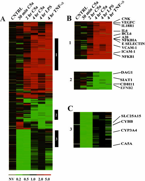
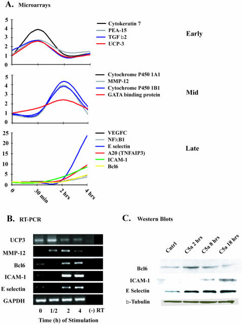
The endothelium plays a critical role in the inflammatory process. The complement activation product, C5a, is known to have proinflammatory effects on the endothelium, but the molecular mechanisms remain unclear. We have used cDNA microarray analysis to assess gene expression in human umbilical vein endothelial cells (HUVECs) that were stimulated with human C5a in vitro. Chip analyses were confirmed by reverse transcriptase-polymerase chain reaction and by Western blot analysis. Gene activation responses were remarkably similar to gene expression patterns of HUVECs stimulated with human tumor necrosis factor-alpha or bacterial lipopolysaccharide. HUVECs stimulated with C5a showed progressive increases in gene expression for cell adhesion molecules (eg, E-selectin, ICAM-1, VCAM-1), cytokines/chemokines, and related receptors (eg, VEGFC, IL-6, IL-18R). Surprisingly, HUVECs showed little evidence for up-regulation of complement-related genes. There were transient increases in gene expression associated with broad functional activities. The three agonists used also caused down-regulation of genes that regulate angiogenesis and drug metabolism. With a single exception, C5a caused little evidence of activation of complement-related genes. These studies indicate that endothelial cells respond robustly to C5a by activation of genes related to progressive expression of cell adherence molecules, and cytokines and chemokines in a manner similar to responses induced by tumor necrosis factor-alpha and lipopolysaccharide.
Figures
References
-
- Madge LA, Poder JS. TNF signaling in vascular endothelial cells. Exp Mol Pathol. 2001;70:317–325. - PubMed
-
- Heumann D, Glauser MP, Calandra T. Molecular basis of host-pathogen interaction in septic shock. Curr Opin Microbiol. 1998;1:49–55. - PubMed
-
- Nooteboom A, Van Der Linden CJ, Hendriks T. Tumor necrosis factor-alpha and interleukin-1beta mediate endothelial permeability induced by lipopolysaccharide-stimulated whole blood. Crit Care Med. 2002;30:2063–2068. - PubMed
-
- Pohlman TH, Harlan JM. Human endothelial cell response to lipopolysaccharide, interleukin-1, and tumor necrosis factor is regulated by protein synthesis. Cell Immunol. 1989;119:41–52. - PubMed
Publication types
MeSH terms
Substances
Grants and funding
LinkOut - more resources
Full Text Sources
Other Literature Sources
Miscellaneous
