

An anti-apoptotic role for galectin-3 in diffuse large B-cell lymphomas
- PMID: 14982843
- PMCID: PMC1614710
- DOI: 10.1016/S0002-9440(10)63177-X
An anti-apoptotic role for galectin-3 in diffuse large B-cell lymphomas
Abstract
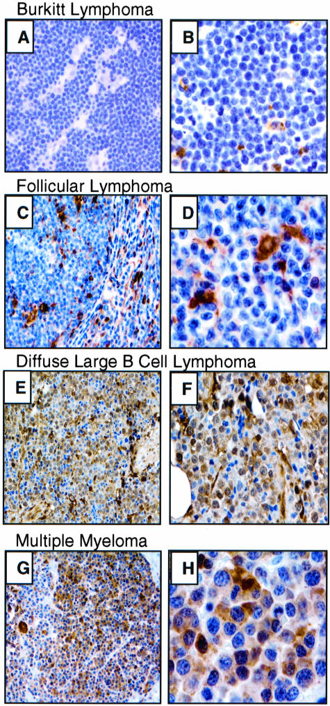
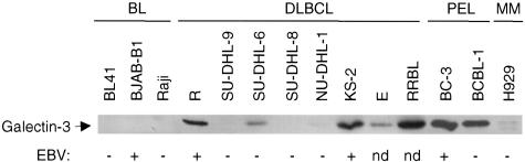
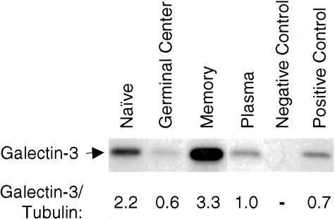
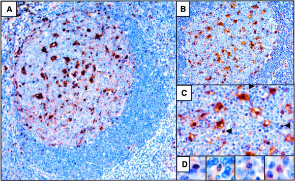
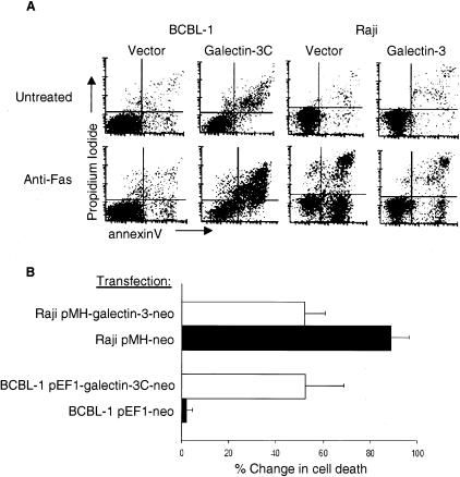
Increased resistance to apoptosis promotes lymphomagenesis with aberrant expression of cell survival proteins such as BCL-2 and c-MYC occurring in distinct lymphoma subtypes. Galectin-3 is an anti-apoptotic protein that protects T cells, macrophages, and breast carcinoma cells from death triggered by a variety of agents. We have found high levels of galectin-3 protein expression in a subset of B-cell neoplasms including diffuse large B-cell lymphoma (DLBCL), primary effusion lymphoma (PEL), and multiple myeloma (MM), in both cell lines and patient samples. However, we failed to detect galectin-3 in Burkitt lymphoma (BL), follicular lymphoma (FL), marginal zone lymphoma (MZL), MALT lymphoma or B-small lymphocytic lymphoma (B-SLL) cell lines or patient samples. To determine whether galectin-3 expression protects B cells from apoptosis, galectin-3-negative BL cells were transfected with a galectin-3 expressing plasmid, which resulted in markedly increased resistance to anti-Fas-induced cell death. In contrast, galectin-3-positive PEL cells transfected with an amino-terminal truncated galectin-3 vector showed increased sensitivity to anti-Fas induced apoptosis. During normal B-cell development, galectin-3 expression was lowest in germinal center and plasma B cells, from which DLBCL, PEL, and MM derive, and highest in long-lived naïve and memory B cells. This pattern of expression suggests that aberrantly increased galectin-3 levels in specific B-cell populations may yield a protective advantage during transformation and/or progression of certain B-cell neoplasms.
Figures
Similar articles
-
T-cell leukemia 1 expression in nodal Epstein-Barr virus-negative diffuse large B-cell lymphoma and primary mediastinal B-cell lymphoma.Hum Pathol. 2010 Sep;41(9):1238-44. doi: 10.1016/j.humpath.2010.01.015. Epub 2010 Apr 10. Hum Pathol. 2010. PMID: 20382409
-
FAS (CD95) mutations are rare in gastric MALT lymphoma but occur more frequently in primary gastric diffuse large B-cell lymphoma.Am J Pathol. 2004 Mar;164(3):1081-9. doi: 10.1016/S0002-9440(10)63195-1. Am J Pathol. 2004. PMID: 14982861 Free PMC article.
-
TCL1 oncogene expression in B cell subsets from lymphoid hyperplasia and distinct classes of B cell lymphoma.Lab Invest. 2001 Apr;81(4):555-64. doi: 10.1038/labinvest.3780264. Lab Invest. 2001. PMID: 11304575
-
Helicobacter pylori and mucosa-associated lymphoid tissue: what's new.Hematology Am Soc Hematol Educ Program. 2013;2013:109-17. doi: 10.1182/asheducation-2013.1.109. Hematology Am Soc Hematol Educ Program. 2013. PMID: 24319171 Review.
-
Expression of apoptosis regulators in germinal centers and germinal center-derived B-cell lymphomas: insight into B-cell lymphomagenesis.Pathol Int. 2007 Jul;57(7):391-7. doi: 10.1111/j.1440-1827.2007.02115.x. Pathol Int. 2007. PMID: 17587238 Review.
Cited by
-
HCV infection and B-cell lymphomagenesis.Adv Hematol. 2011;2011:835314. doi: 10.1155/2011/835314. Epub 2011 Jul 20. Adv Hematol. 2011. PMID: 21789042 Free PMC article.
-
Multifaceted roles of Galectins: from carbohydrate binding to targeted cancer therapy.Biomark Res. 2025 Mar 25;13(1):49. doi: 10.1186/s40364-025-00759-1. Biomark Res. 2025. PMID: 40134029 Free PMC article. Review.
-
GCS-100, a novel galectin-3 antagonist, modulates MCL-1, NOXA, and cell cycle to induce myeloma cell death.Blood. 2010 May 13;115(19):3939-48. doi: 10.1182/blood-2009-10-251660. Epub 2010 Feb 26. Blood. 2010. PMID: 20190189 Free PMC article.
-
Effect of Meloxicam on the Proliferation and Apoptosis of the Raji Cell Line: an In Vitro Study.Int J Dent. 2022 Jul 6;2022:9579326. doi: 10.1155/2022/9579326. eCollection 2022. Int J Dent. 2022. PMID: 35847347 Free PMC article.
-
Galectin Family Members: Emerging Novel Targets for Lymphoma Therapy?Front Oncol. 2022 May 23;12:889034. doi: 10.3389/fonc.2022.889034. eCollection 2022. Front Oncol. 2022. PMID: 35677161 Free PMC article. Review.
References
-
- Chow DA, Greenberg AH. The generation of tumor heterogeneity in vivo. Int J Cancer. 1980;25:261–265. - PubMed
-
- Torres J., Jr Increased sister-chromatid exchanges accompany serial transplantation of murine S49.1 lymphoma. Mutat Res. 1982;105:337–342. - PubMed
-
- French SW, Dawson DW, Miner MD, Doerr JR, Malone CS, Wall R, Teitell MA. DNA methylation profiling: a new tool for evaluating hematologic malignancies. Clin Immunol. 2002;103:217–230. - PubMed
-
- Vose JM. Current approaches to the management of non-Hodgkin’s lymphoma. Semin Oncol. 1998;25:483–491. - PubMed
-
- Staudt LM. Gene expression profiling of lymphoid malignancies. Annu Rev Med. 2002;53:303–318. - PubMed
Publication types
MeSH terms
Substances
Grants and funding
LinkOut - more resources
Full Text Sources
Other Literature Sources
Research Materials
Miscellaneous
