. 2004 Mar;164(3):915-24.
doi: 10.1016/S0002-9440(10)63179-3.

Intervertebral disc degeneration: the role of the mitochondrial pathway in annulus fibrosus cell apoptosis induced by overload

Affiliations

Intervertebral disc degeneration: the role of the mitochondrial pathway in annulus fibrosus cell apoptosis induced by overload

François Rannou et al. Am J Pathol. 2004 Mar.

Abstract

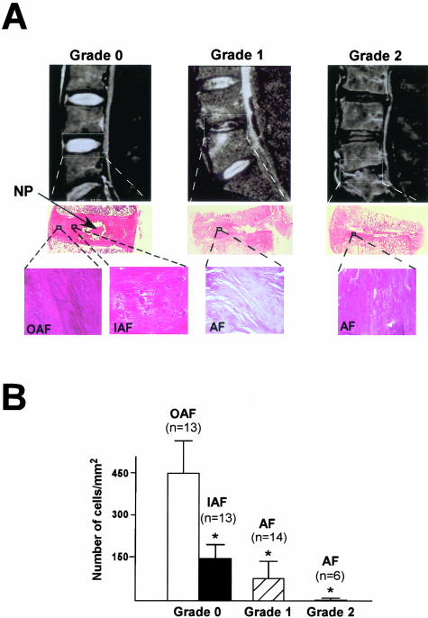
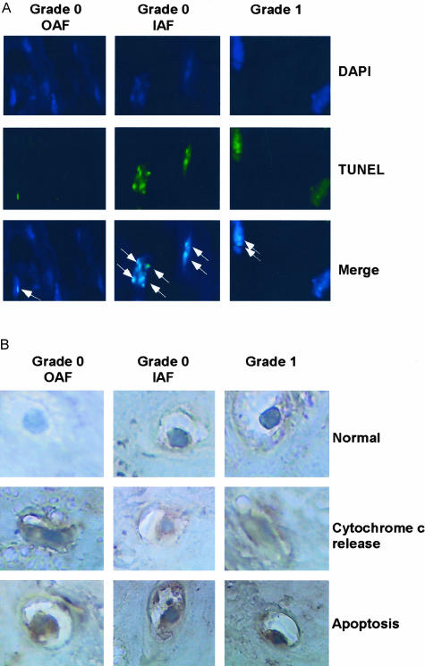
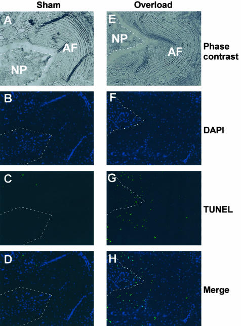
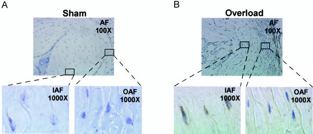
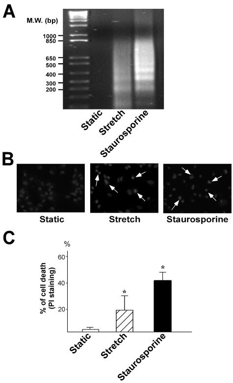
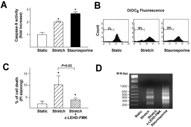
Degeneration of the intervertebral disk (IVD) is a major pathological process implicated in low back pain and is a prerequisite to disk herniation. Although mechanical stress is an important modulator of the degeneration, the underlying molecular mechanism remains unclear. The association of human IVD degeneration, assessed by magnetic resonance imaging, with annulus fibrosus cell apoptosis and anti-cytochrome c staining revealed that the activation of the mitochondria-dependent apoptosome was a major event in the degeneration process. Mouse models of IVD degeneration were used to investigate the role of the mechanical stress in this process. The application of mechanical overload (1.3 MPa) for 24 hours induced annulus fibrosus cell apoptosis and led to severe degeneration of the mouse disks. Immunostaining revealed cytochrome c release but not Fas-L generation. The role of the caspase-9-dependent mitochondrial pathway in annulus fibrosus cell apoptosis induced by overload was investigated further with the use of cultured rabbit IVD cells in a stretch device. Mechanical overload (15% area change) induced apoptosis with increased caspase-9 activity and decreased mitochondrial membrane potential. Furthermore, Z-LEHD-FMK, a caspase-9 inhibitor, but not Z-IETD-FMK, a caspase-8 inhibitor, attenuated the overload-induced apoptosis. Our results from human samples, mouse models, and annulus fibrosus culture experiments demonstrate that the mechanical overload-induced IVD degeneration is mediated through the mitochondrial apoptotic pathway in IVD cells.


Figures

Figure 1
Figure 1
Cell death in human IVD degeneration. A: The top panels are representative MRIs of human lumbar spines. The selected areas represent the IVD categorized as grade 0 (no degeneration), grade 1 (moderate degeneration), and grade 2 (severe degeneration). The center panels reveal the hematoxylin and eosin staining of midsagittal sections of the selected IVD. NP denotes nucleus pulposus. Bottom panels are representative views of the outer annulus fibrosus (OAF), inner annulus fibrosus (IAF) of grade 0 IVDs, and whole AF of grades 1 and 2 IVDs. Magnification: ×100. B: Average number of cells per square millimeter from three views of each IVD at ×100 magnification. * indicates P < 0.05 relative to OAF. n, number of IVDs examined.
Figure 2
Figure 2
Activation of the MDA in human IVD degeneration. A: DAPI (top) and TUNEL (center) staining of OAF, IAF of grade 0 IVDs, and AF of grade 1 disks. Bottom panels represent the merged images of DAPI and TUNEL staining. Arrows indicate the condensed and fragmented nuclei of apoptotic cells. Data are representative views from various IVDs. Magnification: ×400. B: The corresponding cytochrome c immunostaining of specimens shown in A. Top panels are normal AF cells (little immunostaining and normal nuclear morphological features). Center panels show cells having a diffusive pattern of immunoreactivity in the cytoplasm with normal nuclear morphological features, indicating the release of cytochrome c. Bottom panels represent cells with robust cytochrome c staining, cytoplasmic condensation, and nuclear fragmentation. Magnification: ×1250.
Figure 3
Figure 3
Apoptosis in mouse model of IVD degeneration induced by mechanical stress. Left panels are representative IVDs from mice receiving sham surgery; right panels are representative IVDs from mice receiving overload at 1.3 MPa by an external compression device for 24 hours. A and E: Microscopic views of the AF and NP of IVDs. B and F: The corresponding DAPI staining. C and G: TUNEL staining on the same section. D and H: Merged images of DAPI and TUNEL staining. Magnification: ×100. Note: the apoptotic AF cells are mainly located in the inner AF.
Figure 4
Figure 4
Activation of the MDA in mouse IVD degeneration induced by mechanical stress. A: A representative IVD from mice receiving sham surgery, whereas B represents an IVD receiving overload with the same mechanical conditions as those in Figure 3. Data shown are cytochrome c immunostaining under different microscopic magnifications: top, ×100; bottom, ×1000. Note: high levels of cytochrome c immunoreactivity is mainly found in the cytosol of inner AF cells of the IVD subjected to mechanical overload.
Figure 5
Figure 5
Cyclic stretch induces IVD cell apoptosis in vitro. Rabbit AF cells were kept as static controls, subjected to stretch at 15% for 24 hours, or treated with 1 μmol/L staurosporine for 10 hours. A: Genomic DNAs isolated from static, stretched, or staurosporine-treated AF cells were subjected to LM-PCR analysis followed by agarose gel electrophoresis. The data represent results of three independent experiments. B: The nuclear chromatin of AF cells was detected by Hoechst 33258 staining and indicates the cells undergoing apoptosis. Arrows indicate condensed and brightly stained nuclei. A representative section is shown at ×200 magnification. C: Quantitative evaluation of apoptosis was assayed by FACS after PI staining. Results are presented as the percentage of cells below the G0/G1 peak (containing hypodiploid DNA) and are expressed as mean ± SD of three independent experiments. * indicates P < 0.05 relative to static controls.
Figure 6
Figure 6
Cyclic stretch-induced apoptosis is initiated by MDA. Rabbit AF cells were kept as static controls (A and B), stretched at 15% for 24 hours, or treated with 1 μmol/L staurosporine or kept as static controls (C and D), stretched at 15% for 24 hours, or treated with 100 μmol/L Z-LEHD-FMK. A: The caspase-9 activity in cell lysates was measured by detecting the cleavage of LEHD-p-NA, determined by use of a colorimetric plate reader at 405 nm. Results are expressed as mean ± SD from three independent experiments. B: ΔΨ was assayed by FACS after DiOC6 staining. The percentage represents the fraction of cells with decreased DiOC6 fluorescence. C: Quantitative evaluation of apoptosis was assayed by FACS after PI staining. Results are presented as the percentage of cells below the G0/G1 peak (containing hypodiploid DNA) and expressed as mean ± SD from three independent experiments. D: DNA fragmentation was examined after LM-PCR analysis of genomic DNA followed by agarose gel electrophoresis. * indicates P < 0.05 relative to static controls.

References

    1. Annunen S, Paassilta P, Lohiniva J, Perala M, Pihlajamaa T, Karppinen J, Tervonen O, Kroger H, Lahde S, Vanharanta H, Ryhanen L, Goring HH, Ott J, Prockop DJ, Ala-Kokko L. An allele of COL9A2 associated with intervertebral disc disease. Science. 1999;285:409–412. - PubMed
    1. Kawaguchi Y, Osada R, Kanamori M, Ishihara H, Ohmori K, Matsui H, Kimura T. Association between an aggrecan gene polymorphism and lumbar disc degeneration. Spine. 1999;24:2456–2460. - PubMed
    1. Hoogendoorn WE, van Poppel MNM, Bongers PM, Koes BW, Bouter LM. Systematic review of psychosocial factors at work and private life as risk factors for back pain. Spine. 2000;25:2114–2125. - PubMed
    1. Paassilta P, Lohiniva J, Goring HH, Perala M, Raina SS, Karppinen J, Hakala M, Palm T, Kroger H, Kaitila I, Vanharanta H, Ott J, Ala-Kokko L. Identification of a novel common genetic risk factor for lumbar disk disease. JAMA. 2001;285:1843–1849. - PubMed
    1. Pincus T, Burton AK, Vogel S, Field AP. A systematic review of psychological factors as predictors of chronicity/disability in prospective cohorts of low back pain. Spine. 2002;27:109–120. - PubMed

Publication types

MeSH terms