. 2004 Mar;164(3):937-46.
doi: 10.1016/S0002-9440(10)63181-1.

Lipoxins inhibit Akt/PKB activation and cell cycle progression in human mesangial cells

Affiliations

Lipoxins inhibit Akt/PKB activation and cell cycle progression in human mesangial cells

Derick Mitchell et al. Am J Pathol. 2004 Mar.

Abstract

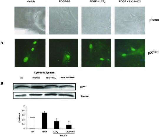
Lipoxins (LX) are endogenously produced eicosanoids with a spectrum of bioactions that suggest anti-inflammatory, pro-resolution roles for these agents. Mesangial cell (MC) proliferation plays a pivotal role in the pathophysiology of glomerular inflammation and is coupled to sclerosis and tubulointerstitial fibrosis. We have previously reported that LXA4 acts through a specific G-protein-coupled-receptor (GPCR) to modulate MC proliferation in response to the proinflammatory mediators LTD4 and platelet-derived growth factor (PDGF). Further investigations revealed that these effects were mediated by modulation of receptor tyrosine kinase activity. Here we have explored the underlying mechanisms and report inhibition of growth factor (PDGF; epithelial growth factor) activation of Akt/PKB by LXA4. LXA4 (10 nmol/L) modulates PDGF-induced (10 ng/ml, 24 hours) decrements in the levels of cyclin kinase inhibitors p21Cip1 and p27Kip1. PDGF-induced increases in CDK2-cyclin E complex formation are also inhibited by LXA4. The potential of LXA4 as an anti-inflammatory therapeutic is compromised by its degradation; this has been circumvented by synthesis of stable analogs. We report that 15-(R/S)-methyl-LXA4 and 16-phenoxy-LXA4 mimic the native compound with respect to modulation of cell proliferation and PDGF-induced changes in cell cycle proteins. In vivo, MC proliferation in response to PDGF is associated with TGFbeta1 production and the subsequent development of renal fibrosis. Here we demonstrate that prolonged (24 to 48 hours) exposure to PDGF is associated with autocrine TGFbeta1 production, which is significantly reduced by LXA4. In aggregate these data demonstrate that LX inhibit PDGF stimulated proliferation via modulation of the PI-3-kinase pathway preventing mitogen-elicited G1-S phase progression and suggest the therapeutic potential of LX as anti-fibrotic agents.


Figures

Figure 1
Figure 1
Inhibition of PDGF and EGF-stimulated MC DNA synthesis by lipoxins. A and B: Subconfluent human MCs were serum restricted in 0.2% FCS medium for 48 hours before stimulation with LXA4 (1 nmol/L), 15-(R/S)-LXA4 (10 pM), 16-phenoxy-LXA4 (10 pM), AG1296 (10 μmol/L) (A), AG1478 (100 nmol/L) (B), for 60 minutes before stimulation with PDGF-BB (10 ng/ml) (A) and EGF (50 ng/ml) (B). Cell proliferation was measured by [3H]-thymidine incorporation after 48 hours. Vehicle (EtOH <0.1%) and 10% FCS were used as negative and positive controls, respectively. Results are expressed as fold/basal cell proliferation. Data represent the mean ± SD of four (A) and two (B) independent experiments performed in triplicate. ** P < 0.01 vs. vehicle, * P < 0.05 vs. PDGF alone (unpaired Student’s t-test). C: Inhibition of serum-stimulated CHOK1-ALXR proliferation by LXA4 and stable synthetic analogs, effect not seen in vector control cell line. CHO K1 cells (± ALXR) were preincubated with LXA4 (1 nmol/L), 15-(R/S)-LXA4 (10 pM), 16-phenoxy-LXA4 (10 pM), for 60 minutes before stimulation with serum-containing media. Cell proliferation was measured by [3H]-thymidine incorporation after 48 hours. Results are expressed as percentage cell proliferation. Data are mean ± SEM of four independent experiments performed in triplicate. * P < 0.05 vs. 10% serum alone (unpaired Student’s t-test).
Figure 2
Figure 2
Akt phosphorylation is modulated by LXA4. Subconfluent cultures of MC were made quiescent in 0.2% FCS medium for 48 hours. MC were then incubated with LXA4 (10 nmol/L, 30′, 60′), LY29002 (3 μmol/L, 30′), LTD4 (10 nmol/L, 30′) (A), 5-HT (10 μmol/L, 30′) (B) before stimulation with PDGF-BB (10 ng/ml, 5′) (A) or EGF (50 ng/ml, 5′) (B). Forty microns of protein were electrophoresed and transferred onto PVDF membrane and immunoblotted for P-Akt (Ser473) or Akt. Results are depicted graphically and represent the mean ± SD for three independent experiments. Values given are fold/basal relative to vehicle-treated cells. * P < 0.001 relative to vehicle, # P < 0.05 relative to PDGF-stimulated cells.
Figure 3
Figure 3
LX regulate PDGF-induced reduction in protein levels of G1 phase. CKI p21Cip1 and p27Kip1 in human MC. A: Subconfluent cultures of human MC were serum restricted in 0.2% FCS medium for 48 hours. Quiescent MC were then stimulated with LXA4 (10 nmol/L, 60′) or vehicle as indicated, before addition of PDGF-BB (10 ng/ml). PDGF-treated MC were co-incubated with TGFβ1 (10 ng/ml) to act as a negative control. MCs were harvested and protein was extracted at 6- and 24-hour intervals poststimulation. Thirty microns protein per lane was resolved by SDS-PAGE and levels of p21Cip1 and p27Kip1 were determined by Western blot. β-actin protein was examined as a loading control. Results are depicted graphically and represent the mean ± SD of three independent experiments. Values given are fold/basal relative to vehicle-treated cells. * P < 0.01 relative to vehicle, # P < 0.05 relative to PDGF-stimulated cells. B: LXA4 stable analogs mimic effect of native LXA4 on PDGF-induced reduction in p21Cip1 and p27Kip1 protein levels. Quiescent MCs were preincubated with LXA4 (10 nmol/L), 15-(R/S)-methyl-LXA4 (10 pM), 16-phenoxy-LXA4 (10 pM) for 60 minutes before treatment with PDGF-BB (10 ng/ml) for 24 hours. Results are depicted graphically and represent the mean ± SD of two independent experiments. Values given are fold/basal relative to vehicle-treated cells.
Figure 4
Figure 4
LXA4 regulates PDGF-induced protein levels of G1 phase CDK2 and cyclin E and CDK2-cyclin E complex formation in human MC. A: Subconfluent cultures of human MC were serum-restricted in 0.2% FCS medium for 48 hours. Quiescent MC were then stimulated with LXA4 (10 nmol/L, 60′) or vehicle as indicated, before addition of PDGF-BB (10 ng/ml). PDGF-treated MC were co-incubated with TGFβ1 (10 ng/ml) to act as a negative control. MCs were harvested and protein was extracted at 6- and 24-hour intervals after stimulation. Thirty microns of protein per lane were size-fractionated by SDS-PAGE and protein expression of cyclin E and CDK2 was determined by Western blot. Expression of β-actin protein was examined as a loading control. Results are depicted graphically and represent the mean ± SD for three independent experiments. Values given are fold/basal relative to vehicle-treated cells. * P < 0.01 relative to vehicle, # P < 0.05 relative to PDGF-stimulated cells. B: The protein from the above conditions was immunoprecipitated with polyclonal antibody to CDK2. The immunoprecipitated lysate was divided in half, separated on an SDS-PAGE, and a Western blot analysis was performed with an antibody to CDK2 and cyclin E. Results are depicted graphically and represent the mean ± SD of two independent experiments. Values given are fold/basal relative to vehicle-treated cells.
Figure 5
Figure 5
PDGF-stimulated TGFβ1 production is attenuated by LXA4. A: Quantitative analysis of TGFβ1 ELISA performed on supernatants of cells at indicated time points. Results are expressed as picograms per microns of MC protein. Data are mean ± SD of seven independent experiments (*P < 0.001 vs. vehicle, # P < 0.05 vs. PDGF alone, unpaired Student’s t-test). B: Subconfluent cultures of human MC were serum restricted in 0.2% FCS medium for 48 hours. Quiescent MC were then stimulated with LXA4 (10 nmol/L, 60′) or vehicle as indicated, before addition of PDGF-BB (10 ng/ml). MCs were harvested and protein was extracted at 6-, 24-, and 48-hour intervals after stimulation. Thirty microns of protein per lane was size-fractionated by SDS-PAGE and protein expression of p27Kip1 was determined by western blot. Expression of β-actin protein was examined as a quantity loading control. C: Western blot analysis at 48 hours poststimulation with above conditions. MCs were stimulated with vehicle, LXA4 (10 nmol/L), PDGF-BB (10 ng/ml) for 48 hours, or pretreated with LXA4 (10 nmol/L) or TGFβ1 monoclonal antibody (1 nmol/L) for 60 minutes before stimulation with PDGF-BB for 48 hours. Results of B and C are depicted graphically and represent the mean ± SD of three independent experiments. Values given are fold/basal relative to vehicle-treated cells. * P < 0.01 relative to vehicle, ** P < 0.05 relative to vehicle, # P < 0.05 relative to PDGF-stimulated cells (24 hours), ## P < 0.05 relative to PDGF-stimulated cells (48 hours).
Figure 6
Figure 6
PDGF increases cytosolic levels of p27Kip1. Influence of LXA4 and LY294002. A: Subcellular localization of p27Kip1 detected by immunocytochemistry. Quiescent MCs on coverslips were incubated with LXA4 (10 nmol/L), LY294002 (3 μmol/L) or vehicle for 30 minutes before stimulation with PDGF (10 ng/ml). At 24 hours, fixed cells were stained with polyclonal antibody against p27Kip1 (green) or phase contrast. B: Cytosolic transition of p27Kip1. Quiescent MCs were incubated with LXA4 (10 nmol/L), LY294002 (3 μmol/L) or vehicle for 30 minutes before stimulation with PDGF (10 ng/ml). After 24 hours, cytosolic and nuclear fractions were extracted. Twenty-five microns of protein per lane were size-fractionated by SDS-PAGE and cytoplasmic levels of p27Kip1 were determined by Western blot. Results are depicted graphically and represent the mean ± SD of three independent experiments. Values given are fold/basal relative to vehicle-treated cells. * P < 0.05 relative to vehicle, # P < 0.05 relative to PDGF-stimulated cells.
Figure 7
Figure 7
Proposed mechanism of LX modulation of PDGF-stimulated MC. proliferation. PDGF stimulation of PDGFRβ in human mesangial cells is coupled to PI-3-kinase and Akt activation, resulting in phosphorylation of p27Kip1 and its translocation from the nucleus, removing the repression on cyclin E-CDK2 activity. In cells treated with LX, inhibition of PDGF receptor phosphorylation is coupled to an attenuation of subsequent Akt phosphorylation and downstream events.

Similar articles

Cited by

References

    1. McMahon BM, Mitchell S, Brady HR, Godson C. Lipoxins: revelations on resolution. Trends Pharmacol Sci. 2001;22:391–395. - PubMed
    1. Serhan CN. Lipoxins and novel aspirin-triggered 15-epi-lipoxins[ATL]: a jungle of cell-cell interactions or a therapeutic opportunity? Prostaglandins. 1997;53:107–137. - PubMed
    1. Levy BD, Clish CB, Schmidt B, Gronert K, Serhan CN. Lipid mediator class switching during acute inflammation: signals in resolution. Nat Med. 2001;2:612–619. - PubMed
    1. Papayianni A, Serhan CN, Phillips ML, Rennke HG, Brady HR. Transcellular biosynthesis of lipoxin A4 during adhesion of platelets and neutrophils in experimental immune complex glomerulonephritis. Kidney Int. 1995:1295–1302. - PubMed
    1. Mayadas TN, Mendrick DL, Brady HR, Tang T, Papayianni A, Assmann KJ, Wagner DD, Hynes RO, Cotran RS. Acute passive anti-glomerular basement membrane nephritis in P-selectin-deficient mice. Kidney Int. 1996;49:1342–1349. - PubMed

Publication types

MeSH terms