Comparative Study
. 2004 Feb 24;32(4):1345-53.
doi: 10.1093/nar/gkh277. Print 2004.

The DNA-binding properties of the ARID-containing subunits of yeast and mammalian SWI/SNF complexes

Affiliations
Comparative Study

The DNA-binding properties of the ARID-containing subunits of yeast and mammalian SWI/SNF complexes

Deborah Wilsker et al. Nucleic Acids Res. .

Abstract

SWI/SNF complexes are ATP-dependent chromatin remodeling complexes that are highly conserved from yeast to human. From yeast to human the complexes contain a subunit with an ARID (A-T-rich interaction domain) DNA-binding domain. In yeast this subunit is SWI1 and in human there are two closely related alternative subunits, p270 and ARID1B. We describe here a comparison of the DNA-binding properties of the yeast and human SWI/SNF ARID-containing subunits. We have determined that SWI1 is an unusual member of the ARID family in both its ARID sequence and in the fact that its DNA-binding affinity is weaker than that of other ARID family members, including its human counterparts, p270 and ARID1B. Sequence analysis and substitution mutagenesis reveals that the weak DNA-binding affinity of the SWI1 ARID is an intrinsic feature of its sequence, arising from specific variations in the major groove interaction site. In addition, this work confirms the finding that p270 binds DNA without regard to sequence specificity, excluding the possibility that the intrinsic role of the ARID is to recruit SWI/SNF complexes to specific promoter sequences. These results emphasize that care must be taken when comparing yeast and higher eukaryotic SWI/SNF complexes in terms of DNA-binding mechanisms.


Figures
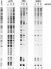
Figure 1
Figure 1
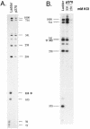
p270 binds DNA non-sequence specifically. λ phage DNA was digested with EcoRI, HindIII and Sau3A1 to generate a large DNA oligonucleotide pool predicted to contain 128 fragments ranging in size from 12 to 2225 bp. The fragments were filled in with [32P]dATP, incubated with GST fusion proteins containing the p270, Dri or MRF2 ARID regions as indicated, pulled down with glutathione beads and analyzed by polyacrylamide gel electrophoresis. Lane 1 shows the unselected pool of DNA fragments. Remaining lanes show the fragments selected in λ DNA binding buffer with increasing KCl concentrations as indicated.
Figure 2
Figure 2
p270 does not bind preferentially to pyrimidine-rich DNA. (A) The pBSII-δ99 plasmid was digested with EcoR1 and Sau3A1 and labeled with [32P]dATP to generate a restriction digest ladder as indicated. The 99 bp pyrimidine-rich fragment is contained within a 110 bp fragment indicated by an asterisk. The restriction fragments were incubated with the p270 GST fusion protein in λ DNA binding buffer at 200 mM KCl. (B) The pBSII-δ99 plasmid was digested with Sau3A1 alone such that the pyrimidine-rich sequence is contained in a 332 bp fragment, indicated by an asterisk. Results from incubations at both 200 and 250 mM KCl are shown.
Figure 3
Figure 3
SWI1 binds DNA non-sequence specifically. The DNA-binding activity of a GST fusion protein containing the ARID region of SWI1 was analyzed as described in Figure 1.
Figure 4
Figure 4
ARID-containing subunits of SWI/SNF complexes in yeast, Drosophila and humans. Similar motifs and domains are apparent between the amino acid sequences of yeast SWI1 (accession no. P09547), Drosophila Osa (accession no. Q8IN94) and human p270 (accession no. NM_006015) and ARID1B (accession no. AF253515). Yellow boxes denote the ARID, vertical gray lines indicate LXXLL motifs (L symbolizes leucine and X is any amino acid). LXXLL motifs frequently serve as association sites for liganded nuclear hormone receptors (33,34). Horizontal blue bars indicate glutamine-rich (Q-rich) regions. Such regions are implicated in transcriptional activation (see for example 35).
Figure 5
Figure 5
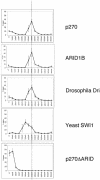
Yeast SWI1 binds DNA poorly compared with other ARID family members, including its human counterparts p270 and ARID1B. In vitro translated [35S]methionine-labeled peptides were applied to a native DNA cellulose column as described under Materials and Methods. Bound protein was eluted stepwise with loading buffer adjusted to contain increasing concentrations of NaCl from 100 to 800 mM, as indicated in the figure. Fractions were separated by SDS–PAGE and the p270 signal in each fraction was quantified by phosphoimaging. The results are plotted as the percentage of signal in each fraction relative to the entire signal recovered. Error bars represent the average deviation. Graphs are aligned for ease of comparison. The dashed line indicates the second 200 mM fraction for reference. The proteins analyzed in this experiment were the respective products of plasmids NE9-B2, KM15, p410, pSWI1.SZ and NNE3ΔARID.
Figure 6
Figure 6
Secondary structure of the ARID. The amino acid sequence of the p270 ARID is aligned according to the Clustal W 1.8 multiple sequence alignment program (36) with the corresponding sequences of SWI1, MRF2 and Dri that were used to generate structural data. The computer-generated alignment was modified slightly to reflect higher level structural data. Residue number 1000 (accession no. NM_006015) is indicated in the p270 sequence for reference. The α-helices of each protein are shaded in yellow and numbered above the alignment. The secondary structure of p270 was determined from the backbone resonance assignments obtained recently (37). H5 and H6 in SWI1 are distinguished by a bend between the two adjacent leucines. The ARID consensus forms six α-helices (H1–H6). p270 has an additional short α-helix at the N-terminus and Dri has an extra α-helix on each end (H0 and H7) formed by sequences outside the consensus. Dri also has a β-sheet in place of Loop 1. While the MRF2 and Dri ARID structures differ in significant features, both structures indicate that H5 and Loop 2 contact the major groove and both structures indicate that sequences between H1 and H2 and sequences just downstream of H6 contact the adjacent minor groove and phosphodiester backbone (18,25,26,27). DNA contact residues identified by NMR in Dri (27) are indicated by red text and underlining. The consensus line shows the residues conserved in more than 50% of the 23 ARID family members of human, Drosophila melanogaster and Saccharomyces cereviseae. Five residues that have proved thus far to be invariant are shown underlined in green.
Figure 7
Figure 7
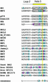
Alignment of the Loop 2 and Helix 5 region of human, Drosophila and yeast ARID family members. The amino acid sequence extending across the Loop 2 and H5 region of all known human, Drosophila and S.cereviseae ARID family members are aligned for comparison. Drosophila Dri and human MRF2 are shown first to help align their defined H5 and Loop 2. The residues that form H5 are boxed where the structure is known for Dri, MRF2, p270 and SWI1. The ARID-containing members of SWI/SNF complexes (SWI1, Drosophila Osa and human p270 and ARID1B) are clustered together. All other mammalian ARID family members are clustered in the third group and the last cluster includes the remaining yeast and Drosophila ARID family members. Basic amino acids, arginine (R), lysine (K) and histidine (H), are shaded blue. Acidic amino acids, aspartic acid (D) and glutamic acid (E), are shaded pink. The invariant tyrosine (Y) residue is shaded yellow. Sequences are aligned according to the invariant tyrosine as well as the highly conserved leucine residues that flank the majority of sequences shown. Dashes are inserted where appropriate to maintain the alignment. The consensus line represents residues conserved in at least 50% of the sequences shown. The blue shaded B in the consensus line represents conservation of basic residues at that position.
Figure 8
Figure 8
Mutants generated in p270 to mimic the yeast SWI1 sequence. The Loop 2 and H5 region of wild-type p270 and SWI1 are shown in the top two lines. Four residues, glycine (G), threonine (T) and two serines (S), in Loop 2 of p270 were deleted to create p270ΔL2. To create the mutant p270ΔL2-DES, an alanine (A) and two lysines (K) were changed to the corresponding SWI1 residues, aspartic acid (D), glutamic acid (E) and serine (S). The substituted positions are indicated by black dots. The invariant tyrosine is shaded yellow for reference.
Figure 9
Figure 9
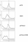
Substitution of SWI1 sequences into p270 is sufficient to make p270 defective for DNA binding. The mutants described in Figure 8 were tested for DNA-binding affinity as described in Figure 4. The wild-type plasmids for p270 and SWI1 (NE9-B2 and pSWI1.SZ) were constructed to generate comparably sized peptides in order to maximize the validity of the comparison. Their elution profiles from Figure 4 are shown again here for ease of comparison. p270ΔL2 and p270ΔL2-DES were constructed in the NE9-B2 background. The dashed line indicates the second 200 mM fraction for reference. Error bars indicate average deviation for at least three experiments. The p270ΔL2-DES elution profile consistently shows two peaks. The reason is not certain, but one possibility is that the accumulated mutations impede proper folding, leading to two populations, one in which structural integrity is severely compromised and another in which the protein has assumed its optimal conformation.

References

    1. Vignali M., Hassan,A.H., Neely,K.E. and Workman,J.L. (2000) ATP-dependent chromatin-remodeling complexes. Mol. Cell. Biol., 20, 1899–1910. - PMC - PubMed
    1. Martens J.A. and Winston,F. (2003) Recent advances in understanding chromatin remodeling by Swi/Snf complexes. Curr. Opin. Genet. Dev., 13, 136–142. - PubMed
    1. Quinn J., Fyrberg,A.M., Ganster,R.W., Schmidt,M.C. and Peterson,C.L. (1996) DNA-binding properties of the yeast SWI/SNF complex. Nature, 379, 844–847. - PubMed
    1. Sengupta S.M., Van Kanegan,M., Persinger,J., Logie,C., Cairns,B.R., Peterson,C.L. and Bartholomew,B. (2001) The interactions of yeast SWI/SNF and RSC with the nucleosome before and after chromatin remodeling. J. Biol. Chem., 276, 12636–12644. - PubMed
    1. Dallas P.B., Cheney,I.W., Liao,D., Bowrin,V., Byam,W., Pacchione,S., Kobayashi,R., Yaciuk,P. and Moran,E. (1998) p300/CREB binding protein-related protein p270 is a component of mammalian SWI/SNF complexes. Mol. Cell. Biol., 18, 3596–3603. - PMC - PubMed

Publication types

MeSH terms

Associated data