. 2004 Feb 24;101(8):2259-64.
doi: 10.1073/pnas.0308762101.

Acetylation of p53 augments its site-specific DNA binding both in vitro and in vivo

Affiliations

Acetylation of p53 augments its site-specific DNA binding both in vitro and in vivo

Jianyuan Luo et al. Proc Natl Acad Sci U S A. .

Abstract

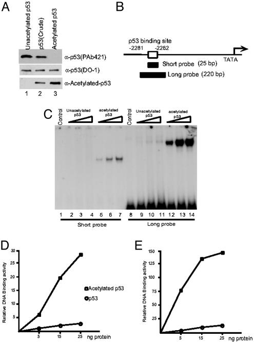
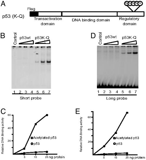
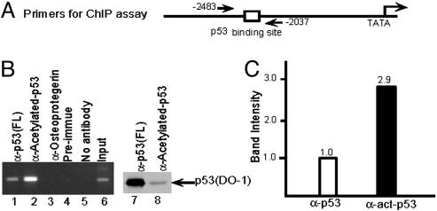
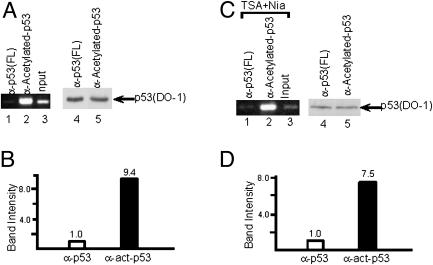
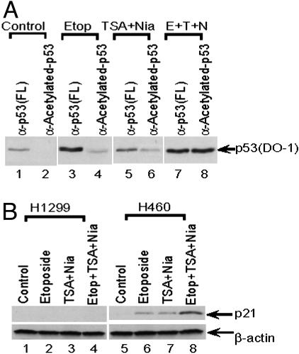
p53 promotes tumor suppression through its ability to function as a transcriptional factor and is activated by posttranslational modifications that include acetylation. Our earlier study demonstrated that p53 acetylation can enhance its sequence-specific DNA binding in vitro, and this notion was later confirmed in several other studies. However, a recent study has reported that in vitro acetylation of p53 fails to stimulate its DNA binding to large DNA fragments, raising an important issue that requires further investigation. Here, we show that unacetylated p53 is able to bind weakly to its consensus site within the context of large DNA fragments, although it completely fails to bind the same site within short oligonucleotide probes. Strikingly, by using highly purified and fully acetylated p53 proteins obtained from cells, we show that acetylation of the C-terminal domain can dramatically enhance site-specific DNA binding on both short oligonucleotide probes and long DNA fragments. Moreover, endogenous p53 apparently can be fully acetylated in response to DNA damage when both histone deacetylase complex 1 (HDAC1)- and Sir2-mediated deacetylation are inhibited, indicating dynamic p53 acetylation and deacetylation events during the DNA damage response. Finally, we also show that acetylation of endogenous p53 indeed significantly augments its ability to bind an endogenous target gene and that p53 acetylation levels correlate well with p53-mediated transcriptional activation in vivo. Thus, our results clarify some of the confusion surrounding acetylation-mediated effects on p53 binding to DNA and suggest that acetylation of p53 in vivo may contribute, at least in part, to its transcriptional activation functions.


Figures

Fig. 1.
Fig. 1.
p53 acetylation strongly enhances its DNA binding activity in vitro.(A) Western blot analysis of purified acetylated p53 (lane 3), unacetylated p53 (lane 1), and a preparation of unresolved acetylated and unacetylated p53 (lane 2) proteins with the antiacetylated p53-specific antibody (Bottom), the DO-1 antibody (Middle), or the PAb421 antibody (Top). (B) Schematic representation of the short and long probes in the p21 promoter region used for EMSA. (C) EMSA assay of unacetylated p53 (lanes 2–4 and 9–11) and acetylated p53 (lanes 5–7 and 12–14) with short (lanes 1–7) and long (lanes 8–14) probes. (D and E) The DNA/protein complexes in C were quantitated by phosphorimaging by using imagequant software (Amersham Biosciences). (D) Results with the short probe. (E) Results with the long probe.
Fig. 2.
Fig. 2.
Lysine to glutamine mutations in the p53 C terminus enhance DNA binding activity in vitro.(A) Schematic representation of the p53 (K-Q) mutant with lysine to glutamine changes at residues 370, 372, 373, 381, and 382. (B and C) EMSA assay of p53 wild type (wt) (lanes 2–4) and p53 (K-Q) mutant (lanes 5–7) with short probe (B). The resulting DNA-protein complexes in B were quantitated by phosphorimaging by using imagequant software (C). (D and F) EMSA assay of p53 wt (lanes 2–4) and p53 (K-Q) mutant (lanes 5–7) with long probe (D). The resulting DNA–protein complexes in D were quantitated by phosphorimaging by using imagequant software (F).
Fig. 3.
Fig. 3.
p53 acetylation strongly enhances its DNA binding activity in vivo.(A) Schematic representation of the primers in p21 promoter region used for ChIP assay. (B) ChIP assay by using anti-p53 (FL) (lane 1), antiacetylated p53 (lane 2), antiosteoprotegerin (lane 3), preimmune (lane 4), and no antibody (lane 5). Western blot shows p53 protein amount in ChIP assay by anti-p53 (FL) (lane 7) and by antiacetylated p53 (lane 8). Cells were treated by etoposide before the ChIP assay. (C) The resulting DNA amounts in ChIP assay by anti-p53 (FL) and antiacetylated p53 (B, lanes 1 and 2) were quantitated by phosphorimaging using imagequant software.
Fig. 4.
Fig. 4.
Acetylation of p53 enhances its DNA binding on endogenous promoters. (A) ChIP assay with equal amounts of immunoprecipitated acetylated and total p53 proteins (lanes 1 and 2). A Western blot with the DO-1 antibody shows the amounts of immunoprecipitated p53 proteins, either by anti-p53 (FL) (lane 4) or by antiacetylated-p53 (lane 5) antibodies, used in the ChIP assays in lanes 1 and 2. Cells were treated by etoposide before the ChIP assay. (B) The resulting DNA amounts in A were quantitated by phosphorimaging by using imagequant software. (C) ChIP assays with anti-p53 (FL) (lane 1) and antiacetylated-p53 (lane 2). Western blot with the DO-1 antibody shows the amounts of immunoprecipitated p53 proteins, either by anti-p53 (FL) (lane 4) or by antiacetylated-p53 (lane 5) antibodies, used in the ChIP assays in lanes 1 and 2. Cells were treated with deacetylase inhibitors TSA and nicotinamide before the ChIP assay. (D) The resulting DNA amounts in C were quantitated by phosphorimaging using imagequant software.
Fig. 5.
Fig. 5.
Endogenous p53 is fully acetylated on DNA damage. (A) Western blot with DO-1 antibody showing the amounts of p53 immunoprecipitated with the indicated p53 antibodies from cells with no treatment (lanes 1 and 2), treatment with etoposide (lanes 3 and 4), treatment with TSA and nicotinamide (lanes 5 and 6), or treatment with etoposide, TSA, and nicotinamide (lanes 7 and 8). (B) Western blot showing p21 levels (Upper) in p53-null H1299 cells (lanes 1–4) or wild-type p53-expressing H460 cells (lanes 5–8) after treatment with the indicated drugs. β-Actin (Lower) was used as a loading control.

References

    1. Levine, A. J. (1997) Cell 88, 323–331. - PubMed
    1. Prives, C. & Hall, P. A. (1999) Pathol. J. 187, 112–126. - PubMed
    1. Vogelstein, B., Lane, D. & Levine, A. J. (2000) Nature 408, 307–310. - PubMed
    1. Hollstein, M., Rice, K., Greenblatt, M. S., Soussi, T., Fuchs, R., Sorlie, T., Hovig, E., Smith-Sorensen, B., Montesano, R. & Harris, C. C. (1994) Nucleic Acids Res. 22, 3551–3555. - PMC - PubMed
    1. Lohrum, M. A. & Vousden, K. H. (1999) Cell Growth Differ. 6, 1162–1168. - PubMed

Publication types

LinkOut - more resources