. 2004 Feb 24;101(8):2392-7.
doi: 10.1073/pnas.0308703100.

Loss of NFAT5 results in renal atrophy and lack of tonicity-responsive gene expression

Affiliations

Loss of NFAT5 results in renal atrophy and lack of tonicity-responsive gene expression

Cristina López-Rodríguez et al. Proc Natl Acad Sci U S A. .

Abstract

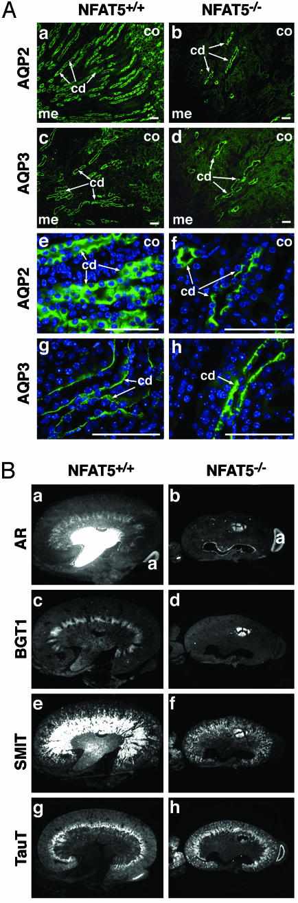
The transcription factor NFAT5/TonEBP, a member of the NFAT/Rel family of transcription factors, has been implicated in diverse cellular responses, including the response to osmotic stress, integrin-dependent cell migration, T cell activation, and the Ras pathway in Drosophila. To clarify the in vivo role of NFAT5, we generated NFAT5-null mice. Homozygous mutants were genetically underrepresented after embryonic day 14.5. Surviving mice manifested a progressive and profound atrophy of the kidney medulla with impaired activation of several osmoprotective genes, including those encoding aldose reductase, Na+/Cl--coupled betaine/gamma-aminobutyric acid transporter, and the Na+/myo-inositol cotransporter. The aldose reductase gene is controlled by a tonicity-responsive enhancer, which was refractory to hypertonic stress in fibroblasts lacking NFAT5, establishing this enhancer as a direct transcriptional target of NFAT5. Our findings demonstrate a central role for NFAT5 as a tonicity-responsive transcription factor required for kidney homeostasis and function.


Figures

Fig. 1.
Fig. 1.
Targeted disruption of the NFAT5 gene. (A) Strategy used to generate the targeted NFAT5 allele. The targeted locus substitutes an inverted Neo cassette for the sixth exon of the NFAT5 gene (first exon of the Rel-homology domain, which encodes the DNA-binding loop), generating a frameshift that introduces a stop codon immediately after the deletion (not shown). B, BamHI; P, PsthAI; A, AvrII; X, XmnI; Bg, BglII. (B) Southern blot analysis from BamHI-restricted genomic DNA with a 5′ probe outside the recombination site identifies the targeted NFAT5 allele in heterozygous mouse crosses. (C) Western blot analysis for NFAT5 protein in lymphocytes from wild-type and NFAT5–/– mice shows complete lack of NFAT5 expression in the mutant. Protein levels of NFAT1 are unaffected in the homozygous mutant (Lower). ns, nonspecific band.
Fig. 2.
Fig. 2.
Growth retardation in NFAT5–/– mice. (A) Comparison of wild-type (Left) and NFAT5–/– (Right) mice demonstrates growth retardation in the mutant at 6 weeks of age. (B) NFAT5–/– mice show a pronounced reduction in body weight compared with wild-type siblings.
Fig. 3.
Fig. 3.
Altered kidney morphology and abnormalities of the renal medulla in NFAT5–/– mice. (A) Histological analysis of hematoxylin/eosin-stained sections from kidneys of 3- to 4-week-old wild-type (a) and NFAT5–/– (b) mice revealed atrophic papillae and a denser morphology of the renal medulla in the mutant (bar = 2 mm). (c and d) In the medulla of the null kidney, the morphology of the inner stripe of the outer medulla is indistinct compared with wild type. Compared with the straight morphology of the tubules in the outer medulla in wild-type kidneys (e), the architecture of the tubules in the kidney medulla in NFAT5–/– mice is curved (f; bar = 100 μm). (g and h) A detailed comparison at a higher magnification of kidneys from wild-type (g) and NFAT5–/– (h) mice revealed that the cells in the renal medulla of NFAT5–/– mice lack normal cuboidal morphology and have a decrease in cytoplasmic volume compared with wild type (bar = 40μm). (i and j) Whereas the medulla is severely affected in NFAT5–/– mice, the histology of the renal cortex is normal (bar = 20 μm). (B) Histological analysis of sections from the kidneys of mature wild-type (a and b) or NFAT5–/– mice (cf). (a, c, and e, bar = 2 μm; b, d, and f, bar = 100 μm). (C) Terminal deoxynucleotidyltransferase-mediated dUTP end-labeling analysis of the renal medulla from wild-type (a) and NFAT5–/– (b) mouse sections indicates increased apoptosis in the mutant. c, cortex; is, inner stripe; m, medulla; om, outer medulla; p, papilla.
Fig. 4.
Fig. 4.
Aberrant renal gene expression in NFAT5–/– mice. (A) Immunostaining of renal tubules. AQP2 (a and b) and AQP3 (c and d) staining reveals abundant expression in collecting ducts (cd) in wild-type kidneys (a and c), but decreased expression in the mutant kidneys (b and d). Higher magnification of AQP2 (ef) and AQP3 (gh) staining of the collecting ducts of wild-type (e and g) and mutant (f and h) kidneys shows predominant cytoplasmic localization in wild-type (e) but apical localization of AQP2 in mutant kidneys (f) and basolateral localization of AQP3 in both wild-type (g) and mutant (h) kidneys. Nuclei were counterstained with 4′,6-diamidino-2-phenylindole in eh. co, cortex; me, medulla. (Bar = 50 μm.) (B) Transcripts encoding AR (a and b), BGT1 (c and d), SMIT (e and f), and TauT (g and h) were detected by in situ hybridization to kidney sections from wild-type (a, c, e, and g) and NFAT5–/– (b, d, f, and h) mice subjected to water restriction for 24 h.
Fig. 5.
Fig. 5.
Down-regulation of AR gene expression in NFAT5–/– mice. (A) Quantification by real-time RT-PCR of levels of AR transcripts induced in wild-type or NFAT5–/– mouse embryo fibroblasts exposed to hypertonic conditions (addition of 100 mM NaCl for 16 h, black bars). Values are normalized to a housekeeping gene (L32). (B) Lack of transcriptional activation of the AR enhancer in osmotically stressed fibroblasts from NFAT5–/– mice. (Upper) Diagram of the enhancer showing the three consensus binding elements for NFAT5. (Lower) Luciferase activity driven by the AR-dependent reporter and normalized to an internal standard of cotransfected Renilla luciferase. A representative experiment of at least three is shown.

References

    1. Burg, M., Kwon, E. & Kultz, D. (1997) Annu. Rev. Physiol. 59, 437–455. - PubMed
    1. Haussinger, D. (1996) Biochem. J. 313, 697–710. - PMC - PubMed
    1. Kwon, H. M. & Handler, J. S. (1995) Curr. Opin. Cell Biol. 7, 465–471. - PubMed
    1. Guyton, A. C. & Hall, J. E. (1996) in Textbook of Medical Physiology (Saunders, Philadelphia), pp. 349–366.
    1. López-Rodríguez, C., Aramburu, J., Rakeman, A. S. & Rao, A. (1999) Proc. Natl. Acad. Sci. USA 96, 7214–7219. - PMC - PubMed

Publication types

Substances