. 2004 Feb 24;101(8):2476-81.
doi: 10.1073/pnas.0308671100.

Adiponectin-induced antiangiogenesis and antitumor activity involve caspase-mediated endothelial cell apoptosis

Affiliations

Adiponectin-induced antiangiogenesis and antitumor activity involve caspase-mediated endothelial cell apoptosis

Ebba Bråkenhielm et al. Proc Natl Acad Sci U S A. .

Abstract

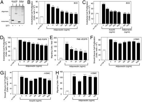
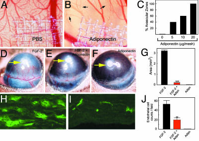
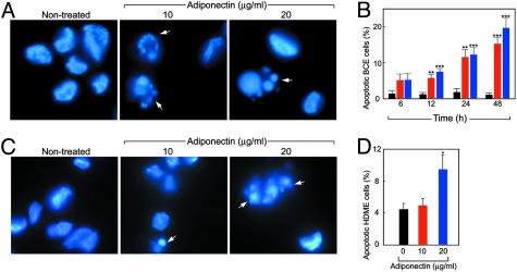
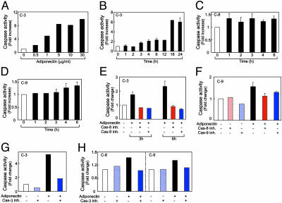
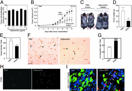
Obesity is a risk factor for the development of many severe human diseases such as cardiovascular disorders, diabetes, and cancer, which are tightly linked to angiogenesis. The adipose tissue produces several growth factors/hormones including leptin, tumor necrosis factor alpha, and adiponectin. It has been found that adiponectin levels are reduced in obesity. Here, we report a unique function of adiponectin as a negative regulator of angiogenesis. In vitro, adiponectin potently inhibits endothelial cell proliferation and migration. In the chick chorioallantoic membrane and the mouse corneal angiogenesis assays, adiponectin remarkably prevents new blood vessel growth. Further, we demonstrate that the antiendothelial mechanisms involve activation of caspase-mediated endothelial cell apoptosis. Adiponectin induces a cascade activation of caspases-8, -9, and -3, which leads to cell death. In a mouse tumor model, adiponectin significantly inhibits primary tumor growth. Impaired tumor growth is associated with decreased neovascularization, leading to significantly increased tumor cell apoptosis. These data demonstrate induction of endothelial apoptosis as an unique mechanism of adiponectin-induced antiangiogenesis. Adiponectin, as a direct endogenous angiogenesis inhibitor, may have therapeutic implications in the treatment of angiogenesis-dependent diseases.


Figures

Fig. 1.
Fig. 1.
Adiponectin inhibits endothelial growth. (A) Recombinant mouse (Acrp30) or human adiponectin (Adipo) proteins (1 μg) were analyzed on a polyacrylamide gel followed by staining with Coomassie brilliant blue. Trimeric/oligomeric (lanes 2 and 4) and monomeric (lanes 1 and 3) forms of adiponectin were detected under nonreducing and reducing/alkylating conditions, respectively. (B and C) FGF-2-stimulated BCE cells were incubated with various concentrations of human or mouse adiponectin. (D) Human adiponectin was incubated with PAE/FGFR-1 cells stimulated with FGF-2. (E) The motility response of PAE/VEGFR-2 cells to VEGF with or without human adiponectin was assayed. FGF-2-stimulated rVSM cells were incubated with human (F) or mouse (G) adiponectin. (H) The motility response of rVSM cells to serum was assayed with or without human adiponectin. Values represent mean number of cells per well (+SEM). *, P < 0.05; **, P < 0.01; ***, P < 0.001.
Fig. 2.
Fig. 2.
Antiangiogenic activity of adiponectin. Disks containing human adiponectin were implanted on the developing CAMs. Microphotographs (×20) on day 8 of a control CAM (A) and an adiponectin-implanted CAM (B;20 μg per disk) are shown. Arrows point to regressing blood vessels. (C) The number of CAMs with avascular zones was quantified at 48 h after implantation. Pellets containing FGF-2 (D), FGF-2/adiponectin (E), or adiponectin alone (F) were implanted into mouse corneal micropockets. Photographs represent ×20 magnification of the mouse eye, and positions of implanted pellets are indicated by arrows. (G) Corneal neovascularization was quantified on day 5 as mean maximal area (+SEM). (H and I) Immunohistochemical labeling of blood vessels in sections of FGF-2-(H) or FGF-2/adiponectin-(I) implanted corneas (×20). (J) The number of microvessels per microscopic field was quantified. **, P < 0.01; ***, P < 0.001.
Fig. 3.
Fig. 3.
Adiponectin induces endothelial apoptosis. (A and B) Apoptotic bodies of adiponectin-treated BCE cells were detected at 24 h by fluorescent microscopy and quantified at different time points. Bars: black, control; red, 10 μg/ml; blue, 20 μg/ml adiponectin. (C and D) Apoptosis of HDME cells after 24-h incubation with adiponectin. Values represent mean percentage of apoptotic cells per total number of cells per field. *, P < 0.05; **, P < 0.01; ***, P < 0.001.
Fig. 4.
Fig. 4.
Activation of endothelial caspase pathways. (A) Dose-dependent activation of caspase-3 in adiponectin-treated BCE cells at 24 h. (B–D) Time course determination of activation of caspase-3-like (B), caspase-8-like (C), and caspase-9-like (D) enzymes in BCE cells treated with 1 μg/ml human adiponectin. Activation of caspase-3 at 3 or 6 h (E and G), caspase-9 at 6 h (F and H), and caspase-8 at 6 h (H) was determined in the presence or absence of specific caspase inhibitors.
Fig. 5.
Fig. 5.
Suppression of tumor growth and induction of apoptosis. (A) T241 tumor cell growth rates in vitro in the presence and absence of mouse adiponectin. (B) Tumor volumes represent mean determinants of treated and control groups (+SEM). (C) Typical examples of tumor-bearing mice of the control or adiponectin-treated groups on day 14. (D) Tumor weights at necropsy. (E) Tumor vascular density was quantified as numbers of vessels per field (×10). (F) Tumor neovascularization was detected by using an anti-CD31 Ab in the adiponectin-treated and control tumors (×20). (Scale bar, 50 μm.) (G) Quantification of TUNEL-positive, apoptotic tumor cells. *, P <0.05; ***, P <0.001. (H) TUNEL staining (green) for visualization of apoptotic cells in tumor sections. (I) Whole-mount staining of tumor blood vessels [×20; green, GFP-T241 tumor cells; blue, CD31-positive tumor vessels; red, TUNEL-positive apoptotic cells (scale bar, 25 μm)]. Arrows indicate CD31/TUNEL double-positive structures.

References

    1. Friedman, J. M. (2000) Nature 404, 632-634. - PubMed
    1. Kopelman, P. G. (2000) Nature 404, 635-643. - PubMed
    1. Guerre-Millo, M. (2002) J. Endocrinol. Invest. 25, 855-861. - PubMed
    1. Frater-Schroder, M., Risau, W., Hallmann, R., Gautschi, P. & Bohlen, P. (1987) Proc. Natl. Acad. Sci. USA 84, 5277-5281. - PMC - PubMed
    1. Claffey, K. P., Wilkison, W. O. & Spiegelman, B. M. (1992) J. Biol. Chem. 267, 16317-16322. - PubMed

Publication types

MeSH terms

LinkOut - more resources