

Coexisting proinflammatory and antioxidative endothelial transcription profiles in a disturbed flow region of the adult porcine aorta
- PMID: 14983035
- PMCID: PMC356976
- DOI: 10.1073/pnas.0305938101
Coexisting proinflammatory and antioxidative endothelial transcription profiles in a disturbed flow region of the adult porcine aorta
Abstract
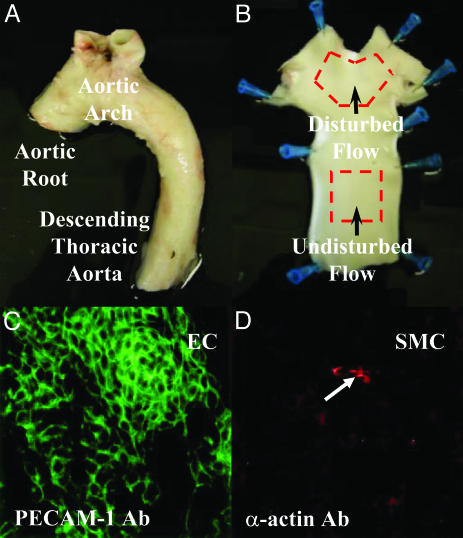
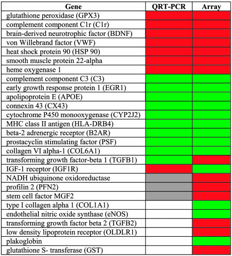
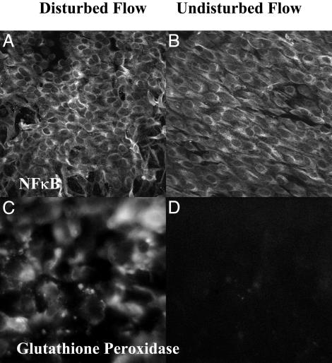
In the arterial circulation, regions of disturbed flow (DF), which are characterized by flow separation and transient vortices, are susceptible to atherogenesis, whereas regions of undisturbed laminar flow (UF) appear protected. Coordinated regulation of gene expression by endothelial cells (EC) may result in differing regional phenotypes that either favor or inhibit atherogenesis. Linearly amplified RNA from freshly isolated EC of DF (inner aortic arch) and UF (descending thoracic aorta) regions of normal adult pigs was used to profile differential gene expression reflecting the steady state in vivo. By using human cDNA arrays, approximately 2,000 putatively differentially expressed genes were identified through false-discovery-rate statistical methods. A sampling of these genes was validated by quantitative real-time PCR and/or immunostaining en face. Biological pathway analysis revealed that in DF there was up-regulation of several broad-acting inflammatory cytokines and receptors, in addition to elements of the NF-kappaB system, which is consistent with a proinflammatory phenotype. However, the NF-kappaB complex was predominantly cytoplasmic (inactive) in both regions, and no significant differences were observed in the expression of key adhesion molecules for inflammatory cells associated with early atherogenesis. Furthermore, there was no histological evidence of inflammation. Protective profiles were observed in DF regions, notably an enhanced antioxidative gene expression. This study provides a public database of regional EC gene expression in a normal animal, implicates hemodynamics as a contributory mechanism to athero-susceptibility, and reveals the coexistence of pro- and antiatherosclerotic transcript profiles in susceptible regions. The introduction of additional risk factors may shift this balance to favor lesion development.
Figures
References
-
- Cornhill, J. F. & Roach, M. R. (1976) Atherosclerosis 23, 489-501. - PubMed
-
- Glagov, S., Zarins, C., Giddens, D. P. & Ku, D. N. (1988) Arch. Pathol. Lab. Med. 112, 1018-1031. - PubMed
-
- Wissler, R. W. (1994) Atherosclerosis 108, S3-S20. - PubMed
-
- Davies, P. F., Reidy, M. A., Goode, T. B. & Bowyer, D. E. (1976) Atherosclerosis 25, 125-130. - PubMed
-
- Faggiotto, A., Ross, R. & Harker, L. (1984) Arteriosclerosis 4, 323-340. - PubMed
Publication types
MeSH terms
Substances
Grants and funding
LinkOut - more resources
Full Text Sources
Molecular Biology Databases
Research Materials
Miscellaneous
