. 2004 Feb 24;101(8):2536-41.
doi: 10.1073/pnas.0307140101.

Potent neutralization of severe acute respiratory syndrome (SARS) coronavirus by a human mAb to S1 protein that blocks receptor association

Affiliations

Potent neutralization of severe acute respiratory syndrome (SARS) coronavirus by a human mAb to S1 protein that blocks receptor association

Jianhua Sui et al. Proc Natl Acad Sci U S A. .

Abstract

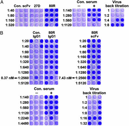
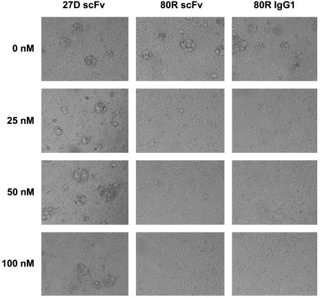
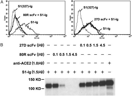
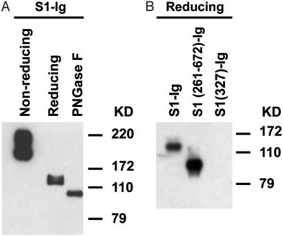
Effective prophylaxis and antiviral therapies are urgently needed in the event of reemergence of the highly contagious and often fatal severe acute respiratory syndrome (SARS) coronavirus (SARS-CoV) infection. We have identified eight recombinant human single-chain variable region fragments (scFvs) against the S1 domain of spike (S) protein of the SARS-CoV from two nonimmune human antibody libraries. One scFv 80R efficiently neutralized SARS-CoV and inhibited syncytia formation between cells expressing the S protein and those expressing the SARS-CoV receptor angiotensin-converting enzyme 2 (ACE2). Mapping of the 80R epitope showed it is located within the N-terminal 261-672 amino acids of S protein and is not glycosylation-dependent. 80R scFv competed with soluble ACE2 for association with the S1 domain and bound S1 with high affinity (equilibrium dissociation constant, Kd=32.3 nM). A human IgG1 form of 80R bound S1 with a 20-fold higher affinity of 1.59 nM comparable to that of ACE2 (Kd=1.70 nM), and neutralized virus 20-fold more efficiently than the 80R scFv. These data suggest that the 80R human monoclonal antibody may be a useful viral entry inhibitor for the emergency prophylaxis and treatment of SARS, and that the ACE2-binding site of S1 could be an attractive target for subunit vaccine and drug development.


Figures

Fig. 1.
Fig. 1.
Microneutralization assay of anti-S1 antibodies on SARS-CoV. (A) Microneutralization assay of anti-S1 scFvs. The positive control was convalescent serum from a SARS patient; the negative control was non-SARS human serum. The names of the scFvs are labeled at the top, and antibody titers are indicated on the left. Neutralizing scFv 80R and only one nonneutralizing scFv 27D (illustrative of the other nonneutralizing scFvs) are shown. Undiluted SARS-CoV (≈37 plaque-forming units) was loaded per well. (B) Comparison of the neutralization activity of 80R scFv and full-length 80R IgG1. The positive and negative control serum samples and the amount of virus used were the same as in A. The titer and concentration of antibodies are labeled on the left.
Fig. 2.
Fig. 2.
Inhibition of syncytia formation by anti-S1 antibodies. Shown is syncytia formation assay with anti-S1 antibodies. 293T cells expressing SARS-CoV S protein were preincubated with the indicated concentrations of anti-S1 scFvs or 80R IgG1 and then mixed with 293T cells expressing ACE2. After culturing for 36 h in the presence of antibodies, dose-dependent inhibition of syncytia formation by 80R scFv, 80R IgG1 was observed and photographed. Representative results are shown.
Fig. 3.
Fig. 3.
80R scFv inhibiting the binding of S1 to ACE2 receptor. (A) Flow cytometry histograms; shown is staining Vero E6 with S1-Ig and flow cytometry analysis. Dotted line, control staining with S1 (327)-Ig; thin line, cells were stained with S1-Ig; bold line, staining with premix of 0.3 μg of S1-Ig and 0.3 μg of 80R scFv (Left) or 27D scFv (Right). (B) scFv competition of S1-Ig binding to ACE2 in immunoprecipitation. Radiolabeled ACE2 was immunoprecipitated by S1-Ig that was preincubated with the indicated amounts of either 27D scFv or 80R scFv. Anti-ACE2 precipitates were used as a positive control. Immunoprecipitates were run on a reducing SDS/PAGE gel and visualized by autoradiography.
Fig. 4.
Fig. 4.
Western blotting of S1-Ig using 80R scFv. Nonreduced, reduced, or deglycosylated samples were subjected to 10% SDS/PAGE gel, transferred to nitrocellulose membrane, detected with anti-S1 80R scFv, and followed by rabbit anti-His-6 Ig and horseradish peroxidase-labeled anti-rabbit IgG. (A) 80R scFv recognized nonreduced S1 much stronger than reduced S1, and there is no further significant decrease of antibody binding to deglycosylated S1 as compared with reduced S1. (B) 80R scFv bound to S1 (261–672)-Ig but did not bind to S1 (327)-Ig. All samples were run under reducing conditions.

References

    1. Ksiazek, T. G., Erdman, D., Goldsmith, C. S., Zaki, S. R., Peret, T., Emery, S., Tong, S., Urbani, C., Comer, J. A., Lim, W., et al. (2003) N. Engl. J. Med. 348, 1953-1966. - PubMed
    1. Kuiken, T., Fouchier, R. A., Schutten, M., Rimmelzwaan, G. F., van Amerongen, G., van Riel, D., Laman, J. D., de Jong, T., van Doornum, G., Lim, W., et al. (2003) Lancet 362, 263-270. - PMC - PubMed
    1. Drosten, C., Gunther, S., Preiser, W., van der Werf, S., Brodt, H.-R., Becker, S., Rabenau, H., Panning, M., Kolesnikova, L., Fouchier, R. A. M., et al. (2003) N. Engl. J. Med. 348, 1967-1976. - PubMed
    1. Holmes, K. V. (2003) N. Engl. J. Med. 348, 1948-1951. - PubMed
    1. Pearson, H., Clarke, T., Abbott, A., Knight, J. & Cyranoski, D. (2003) Nature 424, 121-126. - PMC - PubMed

Publication types

MeSH terms