. 2003 Fall;8(3):272-86.
doi: 10.1379/1466-1268(2003)008<0272:sehiaf>2.0.co;2.

Stress-induced extracellular Hsp72 is a functionally significant danger signal to the immune system

Affiliations

Stress-induced extracellular Hsp72 is a functionally significant danger signal to the immune system

Jay Campisi et al. Cell Stress Chaperones. 2003 Fall.

Abstract

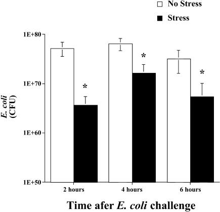
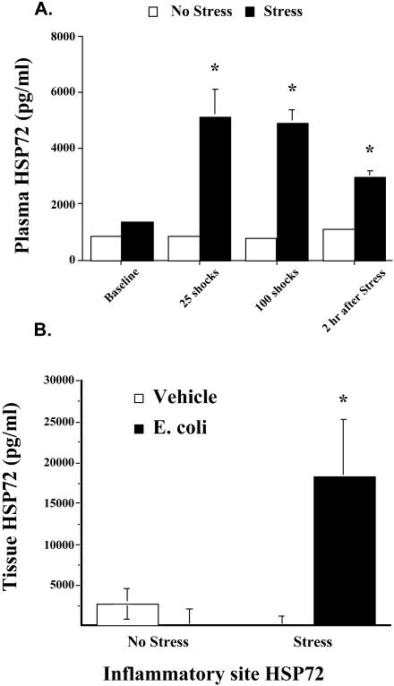
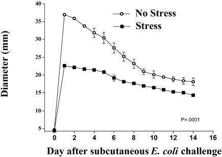
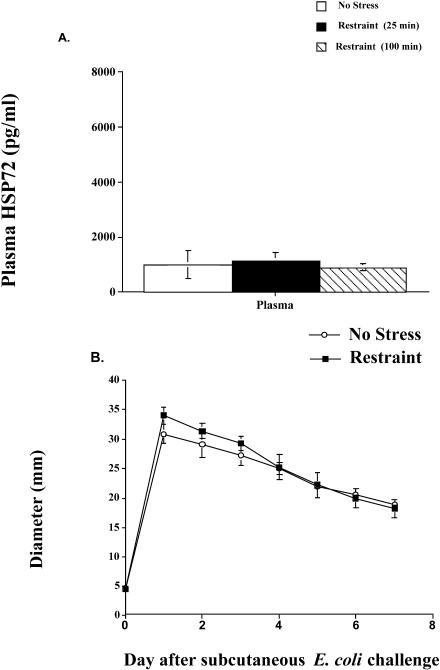
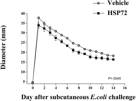
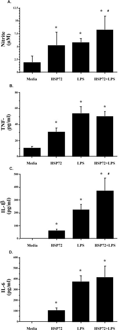
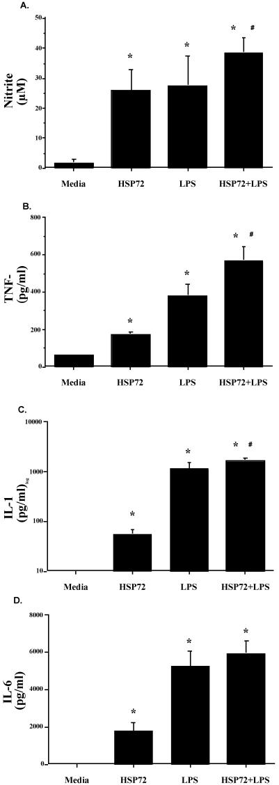
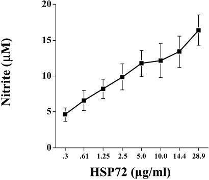
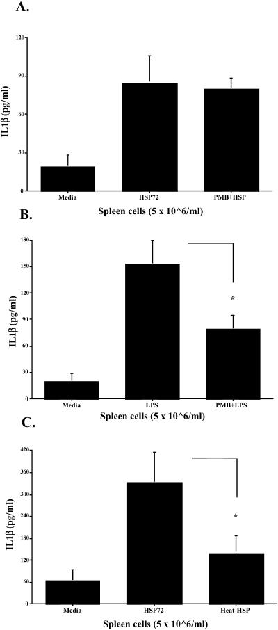
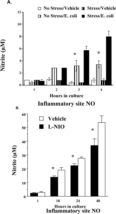
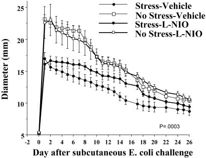
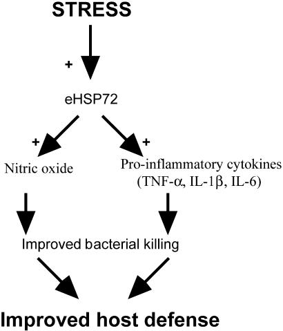
Extracellular heat-shock proteins (eHsp) such as those belonging to the 70-kDa family of Hsp (eg, Hsp72) have been hypothesized to act as a "danger signal" to immune cells, promote immune responses, and improve host defense. The current study tested this hypothesis. Adult male F344 rats were exposed to an acute laboratory stressor (100, 5-second, 1.6-mA inescapable tail shocks) and challenged with Escherichia coli. The number of colony-forming units (CFU) of bacteria at the site of injection, the levels of eHsp72, the immune response to eHsp72 and E. coli-derived lipopolysaccharide (LPS), and the amount of time required to recover from in vivo bacterial challenge were measured. CFUs were reduced 2, 4, and 6 hours after injection of E. coli in rats exposed to stress. Rats exposed to stress had elevated eHsp72 that was elevated rapidly (25 minutes) and remained elevated in the circulation and at the inflammatory site (2 hours after stressor termination). Both stressor exposure and eHsp72 administration in the absence of stress resulted in a facilitated pattern of recovery after bacterial inflammation induced by subcutaneous E. coli injection. Rats exposed to acute restraint (100 minutes) did not demonstrate elevated circulating eHsp72 or a facilitated pattern of recovery after bacterial challenge. In vitro stimulation of rat splenocytes and macrophages with eHsp72 elevated nitric oxide (NO), tumor necrosis factor alpha (TNF-alpha), interleukin (IL)-1beta, and IL-6, and this effect was specific to eHsp72 because it was not diminished by polymyxin B and was reduced by earlier heat-denature treatment. Stimulation of cells with eHsp72 combined with LPS resulted in a greater NO and cytokine response than that observed after stimulation with eHsp72 or LPS alone. In vivo, at the inflammatory site, the bacterial-induced NO response was potentiated by stress, and NO inhibition (L-NIO) reduced the stress-induced facilitation but had no effect on the control kinetics of bacterial inflammation recovery. Thus, these results lend support to the hypothesis that intense stressor exposure increases eHsp72, which acts as a danger signal to potentiate the NO response to bacterial challenge and facilitate recovery from bacterial inflammation.


Figures

Fig 1.
Fig 1.
Adult male F344 rats (n = 7/group/time point) either were exposed to 100, 5-second, 1.6-mA inescapable tail shocks (stress) or remained in their home cages (no stress). Immediately after stressor termination, all rats were injected subcutaneously with live E coli (∼2.5 × 10⁹ colony-forming units [CFU]). Rats were sacrificed 2, 4, or 6 hours after bacterial challenge, and inflammatory sites, previously inoculated with E coli, were excised. Two sites were collected for each rat and combined. Inflammatory sites were homogenized, plated, and quantified. No viable bacteria were recovered from control skin sites. The bacterial counts (CFU) are presented in scientific nomenclature on a log10 scale. All figures represent mean ± standard error of the mean. Stress vs no stress, F-PLSD *P < 0.05
Fig 2.
Fig 2.
(A) Adult male F344 rats (n = 4/group) either were exposed to 100, 5-second, 1.6-mA inescapable tail shocks (stress) or remained in their home cages (no stress). Blood samples were taken the day before the experimental procedure (baseline), after 25 tail shocks (25 minutes), after 100 tail shocks (100 minutes), and 2 hours after stressor termination. Time-matched blood samples were taken from nonstressed home cage controls animals, and eHsp72 was measured. (B) Rats (n = 16) either were exposed to inescapable tail shocks (stress) or remained in their home cages (no stress). Immediately after stressor termination, rats (n = 4/group) were injected either with E coli or with saline (vehicle). Two hours after E coli challenge and stress or no-stress treatment, rats were sacrificed, inflammatory tissue was harvested and placed in culture, and eHsp72 was measured. All figures represent mean ± standard error of the mean. (A) Stress vs no stress, F-PLSD *P < 0.05. (B) Stress (E coli) vs stress (vehicle), F-PLSD *P < 0.05
Fig 3.
Fig 3.
Adult male F344 rats (n = 4/group) either were exposed to 100, 5-second, 1.6-mA inescapable tail shocks (stress) or remained in their home cages (no stress). Immediately after stressor termination, all rats were injected subcutaneously with live E coli (∼2.5 × 10⁹ colony-forming units). Inflammation was measured daily. Group (stress vs no stress) by time interaction, P = 0.0001
Fig 4.
Fig 4.
(A) Adult male F344 rats (n = 5/group) either were exposed to an acute restraint procedure (restraint) or remained in their home cages (no stress). Blood samples were taken after 25 and 100 minutes of restraint. Time-matched blood samples were taken from nonstressed home cage controls (no stress) animals, and eHsp72 was measured. (B) Rats (n = 5/group) either were exposed to 100 minutes of restraint (restraint) or remained in their home cages (no stress). Immediately after restraint, all rats were injected subcutaneously with live E coli (∼2.5 × 10⁹ colony-forming units). Inflammation was measured daily
Fig 5.
Fig 5.
Adult male F344 rats (n = 5 or 6/group) were subcutaneously injected with either 0 μg or 20 μg of Hsp72 and E coli (∼2.5 × 10⁸ colony-forming units) in Freund incomplete adjuvant. Inflammation diameters were recorded daily. Group (vehicle vs Hsp72) by time interaction, P = 0.0045
Fig 6.
Fig 6.
Adult male F344 rats (n = 12) were sacrificed, and spleens were aseptically harvested. Spleen cells (5 × 10⁶ cells/mL, 300 μL/well) were cultured in the presence of 10 μg/mL of Hsp72 protein, 50 μg/mL of lipopolysaccharide (LPS), 10 μg/mL of Hsp72 protein combined with 50 μg/mL of LPS, or an equal volume of culture media. After incubation for 24 hours, (A) nitrite, (B) tumor necrosis factor–α, (C) interleukin (IL)-1β, and (D) IL-6 were measured in the culture supernatants. All figures represent mean ± standard error of the mean. (A–D) Stimulation (Hsp72, LPS, Hsp72 + LPS) vs media, F-PLSD *P < 0.05; Hsp72 + LPS vs Hsp72 or LPS alone, F-PLSD #P < 0.05
Fig 7.
Fig 7.
Adult male F344 rats (n = 12) were sacrificed, and peritoneal cells were aseptically harvested. Peritoneal cavity cells (1 × 10⁶ cells/mL, 300 μL/well) were cultured in the presence of 10 μg/mL of Hsp72 protein, 50 μg/mL of lipopolysaccharide (LPS), 10 μg/mL of Hsp72 protein combined with 50 μg/mL of LPS, or an equal volume of culture media. After incubation for 24 hours, (A) nitrite, (B) tumor necrosis factor–α, (C) interleukin (IL)-1β, and (D) IL-6 were measured in the culture supernatants. All figures represent mean ± standard error of the mean. (A–D) Stimulation (Hsp72, LPS, Hsp72 + LPS) vs media, F-PLSD *P < 0.05; Hsp72 + LPS vs Hsp72 or LPS alone, F-PLSD #P < 0.05
Fig 8.
Fig 8.
Adult male F344 rats (n = 6) were sacrificed, and spleens were aseptically harvested. Spleen cells (5 × 10⁶ cells/mL, 300 μL/well) were cultured in the presence of 0.3–28.9 μg/mL of Hsp72 or vehicle. After incubation for 24 hours, culture supernatant nitrite was measured. All figures represent mean ± standard error of the mean
Fig 9.
Fig 9.
Adult male F344 rats (n = 12) were sacrificed, and splenocytes were aseptically harvested. Spleen cells (5 × 10⁶ cells/mL, 300 μL/well) were cultured for 24 hours with the after-stimulants: (A) culture media (media), 10 μg/mL of Hsp72 protein (Hsp72) or 100 μg/mL of polymyxin B (PMB) + 10 μg/mL of Hsp72 (PMB + Hsp); (B) culture media (media), 50 μg/mL of lipopolysaccharide (LPS) or 100 μg/mL of PMB + 50 μg/mL of LPS (PMB + LPS); or (C) culture media (media), 10 μg/mL of Hsp72 protein (Hsp72) or 10 μg/mL of Hsp72 protein that was heat treated at 100°C for 90 minutes (heat-Hsp). Interleukin 1-β levels were measured from culture supernatants using enzyme-linked immunosorbent assay. All figures represent mean ± standard error of the mean. (B) LPS vs PMB + LPS, F-PLSD *P < 0.05. (C) Hsp72 vs heat-Hsp, F-PLSD *P < 0.05
Fig 10.
Fig 10.
(A) Adult male F344 rats (n = 21) either were exposed to 100, 5-second, 1.6-mA inescapable tail shocks (stress) or remained in their home cages (no stress). Immediately after stressor termination, rats (n = 4–6/group) were injected with either E coli (∼2.5 × 10⁹ colony-forming units [CFU]) or saline (vehicle). Two hours after stressor termination, rats were sacrificed, and inflammatory sites were aseptically dissected, cut into smaller sections, put into 2 mL of Hanks media, and placed in culture. Nitrite was measured from the supernatants after 1, 2, 3, or 4 hours in culture. (B) Unstressed rats (n = 12) were injected with E coli (∼2.5 × 10⁹ CFU). After 1 hour, rats (n = 6/group) were injected again with either l-N5-(1-iminoethyl)-ornithine dihydrochloride (L-NIO) or saline (vehicle) into the inflammatory site. Rats were sacrificed 1 hour after L-NIO injection, and their inflammatory site was aseptically dissected and placed in culture. Supernatants were collected, and nitrite was measured after 1, 18, 24, and 48 hours in culture. All figures represent mean ± standard error of the mean. (A) Stress (E coli) vs no stress (E coli), F-PLSD *P < 0.05. (B) L-NIO vs vehicle, F-PLSD *P < 0.05
Fig 11.
Fig 11.
Adult, male F344 rats (n = 22) either were exposed to 100, 5-second, 1.6-mA inescapable tail shocks (stress) or remained in their home cages (no stress). Immediately after stressor termination, all rats were injected with Escherichia coli (∼2.5 × 10⁹ colony-forming units). Rats (n = 4–6/group) were then injected with equal volumes of either l-N5-(1-iminoethyl)-ornithine dihydrochloride (L-NIO) or vehicle at 0, 30, 60, 90, 120, 150, and 180 minutes after the E coli injection. Inflammation measurements were taken daily. All figures represent mean ± standard error of the mean. Group (stress [vehicle], stress [L-NIO], no stress [vehicle], no stress [L-NIO]) by time interaction, P = 0.0003
Fig 12.
Fig 12.
Schematic of how stress-induced extracellular Hsp72 (eHsp72) could be a functionally significant danger signal to the immune system. Acute stress increases eHsp72 that accumulates at the site of inflammation and facilitates bacterial-stimulated NO and cytokines released by macrophages or neutrophils (or both). Potentiated NO and proinflammatory cytokines facilitate bacterial killing, resulting in faster inflammation resolution and improved host defense

References

    1. Ali H, Haribabu B, Richardson RM, Snyderman R. Mechanisms of inflammation and leukocyte activation. Med Clin North Am. 1997;81:1–28. - PubMed
    1. Asea A, Kraeft SK, Kurt-Jones EA, Stevenson MA, Chen LB, Finberg RW, Koo GC, Calderwood SK. HSP70 stimulates cytokine production through a CD14-dependant pathway, demonstrating its dual role as a chaperone and cytokine. Nat Med. 2000;6:435–442. - PubMed
    1. Asea A, Rehli M, Kabingu E, Boch JA, Bare O, Auron PE, Stevenson MA, Calderwood SK. Novel signal transduction pathway utilized by extracellular HSP70: role of toll-like receptor (TLR) 2 and TLR4. J Biol Chem. 2002;277:15028–15034. - PubMed
    1. Basu S, Binder RJ, Ramalingam T, Srivastava PK. CD91 is a common receptor for heat shock proteins gp96, hsp90, hsp70, and calreticulin. Immunity. 2001;14:303–313. - PubMed
    1. Basu S, Binder RJ, Suto R, Anderson KM, Srivastava PK. Necrotic but not apoptotic cell death releases heat shock proteins, which deliver a partial maturation signal to dendritic cells and activate the NF-kappa B pathway. Int Immunol. 2000;12:1539–1546. - PubMed

Publication types

LinkOut - more resources