. 2004 Feb 25;24(8):1924-35.
doi: 10.1523/JNEUROSCI.4470-03.2004.

The nitric oxide-guanylyl cyclase signaling pathway modulates membrane activity States and electrophysiological properties of striatal medium spiny neurons recorded in vivo

Affiliations

The nitric oxide-guanylyl cyclase signaling pathway modulates membrane activity States and electrophysiological properties of striatal medium spiny neurons recorded in vivo

Anthony R West et al. J Neurosci. .

Abstract

Nitric oxide (NO)-releasing interneurons are believed to regulate the activity of striatal medium spiny neurons (MSNs) that contain the NO effector enzyme guanylyl cyclase (GC). The involvement of NO-GC signaling in modulating steady-state membrane activity of striatal MSNs was examined using in vivo intracellular recordings in rats. Intrastriatal infusion of a neuronal NO synthase inhibitor or a NO scavenger via reverse microdialysis consistently decreased the amplitude of spontaneously occurring depolarized plateau potentials (up events). Intrastriatal infusion of a NO scavenger also decreased the amplitude of EPSPs evoked during electrical stimulation of the orbital prefrontal cortex. The effect of the NO scavenger on spontaneous up events was partially reversed by coperfusion with a cell-permeable cGMP analog. Intracellular injection of MSNs with a soluble GC inhibitor resulted in large decreases in the following: (1) spontaneous up-event amplitude, (2) responsiveness to depolarizing current, (3) action potential amplitude, and (4) input resistance. These effects were partially reversed by coinjection of cGMP. Conversely, intracellular injection of a phosphodiesterase inhibitor increased MSN neuron membrane excitability. These results indicate that, in the intact animal, the NO signaling pathway exerts a powerful tonic modulatory influence over the membrane activity of striatal MSNs via the activation of GC and stimulation of cGMP production.


Figures
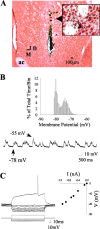
Figure 1.
Figure 1.
Intracellular recordings from striatal neurons located proximal to the microdialysis probe. A, Coronal section (5×) of the striatum demonstrating the location of a striatal neuron (arrow) labeled after intracellular biocytin injection (enlarged to 40× in the inset). Note that the neuron was located proximal (50-100 μm) to the active zone of the microdialysis probe (extends dorsally 4 mm from the termination point of the probe track). ac, Anterior commissure; D, dorsal; M, medial. B, Intracellular recordings from the striatal neuron labeled in A revealed that this cell did not fire spontaneously but did exhibit membrane activity characterized by rapid and spontaneous transitions from a hyperpolarized state to a depolarized plateau. Inset, Example of a time interval plot of membrane potential activity (30 sec recordings sampled at 10 kHz) recorded from the neuron shown in A. C, Left, In the same cell, intracellular injection of 150-msec-duration constant hyperpolarizing current pulses (bottom traces) induced deflections in the membrane potential (top traces). Right, A plot of the steady-state voltage deflections against the current pulse amplitudes taken from recordings shown on the left, revealing an input resistance of 18.3 MΩ for this neuron.
Figure 2.
Figure 2.
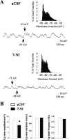
Intrastriatal infusion of the neuronal NOS inhibitor 7-NI decreases the amplitude of spontaneous up events in vivo. A, Top, During aCSF (vehicle) infusion, the majority of striatal neurons exhibited rapid spontaneous shifts in steady-state membrane potential and bimodal membrane potential distributions (inset, 30 sec recordings sampled at 10 kHz) in the absence of spontaneous spike discharge (top trace). Bottom, All of the neurons (n = 4) recorded during local 7-NI infusion (300 μm; 10-60 min) still exhibited up- and down-state activity (bottom trace); however, a significant decrease in the up-state amplitude was observed. Arrows indicate the membrane potential at its maximal depolarized and hyperpolarized levels. B, Left, The mean ± SEM up-state amplitude was reduced after intrastriatal 7-NI infusion (n = 4; *p < 0.05; paired t test). The mean ± SEM up-state duration (center) and frequency of occurrence (right) was not affected by intrastriatal 7-NI infusion (n = 4; p > 0.05; paired t test).
Figure 3.
Figure 3.
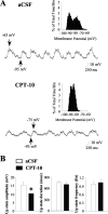
Intrastriatal infusion of the NO scavenger CPT-10 decreases the amplitude of up events. A, Top, During aCSF (vehicle) infusion, the majority of striatal neurons exhibited rapid spontaneous shifts in steady-state membrane potential and bimodal membrane potential distributions (inset, 30 sec recordings sampled at 10 kHz) in the absence of spontaneous spike discharge (top trace). Bottom, All of the neurons (n = 5) recorded during local CPT-10 infusion (1 mm; 5-35 min) still exhibited up- and down-state activity (bottom trace); however, a significant decrease in the up-state amplitude was observed. Arrows indicate the membrane potential at its maximal depolarized and hyperpolarized levels. B, Left, The mean ± SEM up-state amplitude was reduced after intrastriatal CPT-10 infusion (n = 5; *p < 0.05; paired t test). Right, The mean ± SEM up-state duration (center) and frequency of occurrence (right) was not affected by intrastriatal CPT-10 infusion (n = 5; p > 0.05; paired t test).
Figure 4.
Figure 4.
Intrastriatal CPT-10 infusion decreases the responsiveness of striatal neurons to electrical stimulation of the orbital prefrontal cortex. A series of single pulses (0.2 Hz) of electrical stimuli were delivered to the oPFC as described in Materials and Methods during intrastriatal aCSF or CPT-10 infusion (1 mm). A, Representative traces of maximal EPSPs evoked in the same striatal neuron during aCSF and CPT-10 infusion. B, The mean amplitude of EPSPs (expressed as percentage of maximal response) evoked by 800 and 1000 μA stimulus intensities was reduced after intrastriatal CPT-10 infusion (n = 5; **p < 0.01; two-way repeated-measures ANOVA with Dunnett's post hoc test). C, The mean duration of EPSPs (expressed as percentage of maximal response) evoked by 1000 μA stimulus intensities was reduced after intrastriatal CPT-10 infusion (n = 5; *p < 0.05; two-way repeated-measures ANOVA with Dunnett's post hoc test). The mean onset latency of evoked EPSPs and membrane potential before stimulation were not significantly different during aCSF or CPT-10 infusion (data not shown). Filled circles indicate the mean ± SEM EPSP amplitude-duration recorded during aCSF (control) infusion. Open squares indicate the mean ± SEM EPSP amplitude-duration after CPT-10 infusion.
Figure 5.
Figure 5.
Intrastriatal infusion of CPT-10 decreases paired-pulse facilitation of EPSPs evoked by electrical stimulation of the orbital prefrontal cortex. A series of paired pulses (0.2 Hz, 1 mA stimulus intensity; 50 or 100 msec interstimulus interval) of electrical stimuli were delivered to the oPFC as described in Materials and Methods during either intrastriatal aCSF or CPT-10 infusion (1 mm). A, Examples of responses evoked by paired-pulse stimulation delivered at 50 (top) and 100 (bottom) msec interstimulus intervals in the same striatal neuron during CPT-10 infusion. B, Examples of paired-pulse facilitation of spike activity evoked during electrical stimulation [delivered at 50 (top) and 100 (bottom) msec interstimulus intervals] of corticostriatal pathways during CPT-10 infusion. The delivery of the first and second pulses of the pair is indicated by the numbers 1 and 2, respectively. Solid arrows indicate the membrane potential before stimulation. C, The mean ± SEM amplitude of EPSPs [expressed as percentage of the amplitude of the first (control) EPSP of the pair] evoked by 1000 μA stimulus intensities was reduced after intrastriatal CPT-10 infusion compared with control cells recorded during aCSF infusion (n = 3-4; *p < 0.05; t test).
Figure 6.
Figure 6.
Intrastriatal infusion of CPT-10 modulates the membrane activity of striatal neurons in manner that is partially reversed by coperfusion with a cell-permeable cGMP analog. Striatal neurons were recorded after intrastriatal infusion of either aCSF (control), the NO scavenger CPT-10 (1 mm), or CPT-10 (1 mm) plus the cell-permeable cGMP analog 8-Br-cGMP (2 mm). A, Left, After aCSF infusion (5 min), striatal neurons (n = 11) exhibited typical rapid spontaneous shifts in steady-state membrane potential. Right, Time interval plots of membrane potential activity recorded from control neurons demonstrated bimodal membrane potential distributions indicative of bistable membrane activity. B, Left, Similar to within-subjects studies, striatal neurons recorded after CPT-10 infusion (5 min) exhibited significantly lower amplitude up events compared with controls. Right, Time interval plots of membrane potential activity recorded from neurons recorded after CPT-10 infusion demonstrated bimodal membrane potential distributions. C, Left, Striatal neurons recorded after CPT-10 and 8-Br-cGMP infusion (5 min) exhibited high amplitude up events and higher rates of spontaneous spike discharge. Right, The membrane potential distribution of neurons recorded after CPT-10 and 8-Br-cGMP infusion was more depolarized than controls (Table 1). D, Left, The mean ± SEM up-state amplitude was reduced after CPT-10 infusion in a manner that was partially reversed by inclusion of 8-Br-cGMP in the perfusion buffer (n = 8-11 cells; *p < 0.05; ANOVA with Dunnett's post hoc test). Right, The mean ± SEM up-state duration was not significantly altered by either drug treatment (n = 8-11 cells; p > 0.05; ANOVA with Dunnett's post hoc test).
Figure 7.
Figure 7.
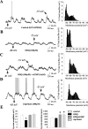
Manipulation of intracellular cGMP levels modulates the membrane activity of striatal neurons. Striatal neurons were recorded after intracellular application (∼5 min) of either vehicle (0.5% DMSO), the GC inhibitor ODQ (100 μm), ODQ plus cGMP (1 mm), or the PDE inhibitor zaprinast (200 μm). A, Left, After vehicle injection, striatal neurons (n = 6) exhibited typical rapid spontaneous shifts in steady-state membrane potential and irregular spontaneous spike discharge. Right, Time interval plots of membrane potential activity recorded from control neurons demonstrated bimodal membrane potential distributions indicative of bistable membrane activity. B, Left, Striatal neurons recorded after ODQ injection (n = 5) exhibited significantly lower amplitude up events compared with vehicle-injected controls and rarely fired action potentials. Right, The depolarized portion of the membrane potential distribution of neurons recorded after ODQ injection was typically shifted leftward (hyperpolarized) compared with controls. C, Left, Striatal neurons recorded after ODQ and cGMP injection rarely fired action potentials but exhibited high amplitude up events with extraordinarily long durations. Right, The membrane potential distribution of neurons recorded after ODQ and cGMP injection was similar to controls, indicating that cGMP partially reversed some of the effects of ODQ. D, Left, Striatal neurons recorded after intracellular injection of zaprinast (n = 5) exhibited high amplitude up events with extraordinarily long durations. Additionally, all of the cells fired action potentials at relatively high rates (0.4-2.2 Hz). Right, The membrane potential distribution of these neurons was typically shifted rightward (depolarized) compared with controls. Arrows indicate the membrane potential at its maximal depolarized and hyperpolarized levels. E, Left, The mean ± SEM up-state amplitude was reduced after intracellular injection of ODQ (n = 5; *p < 0.05; ANOVA with Dunnett's post hoc test). Right, The mean ± SEM up-state duration was significantly prolonged during cGMP/ODQ and zaprinast injections (n = 5; *p < 0.05; ANOVA with Dunnett's post hoc test).
Figure 8.
Figure 8.
Intracellular injection of the guanylyl cyclase inhibitor ODQ decreased the responsiveness of striatal neurons to intracellular current injection. A, Left column, Response of a single cell to increasing amplitudes of intracellular current injected after vehicle (0.5% DMSO) injection. Middle column, Decreased responsiveness of a typical single cell to depolarizing current pulses injected after ODQ injection (∼5-6 min). Note that the current amplitude required to reach threshold (rheobase) is significantly larger (1.15 nA) than the representative control (0.20 nA). Right column, Partial reversal of the ODQ effect after coinjection with cGMP (∼5-6 min). Bottom traces indicate current injection steps. Top traces indicate the voltage response. The membrane potential before current injection is indicated below the voltage trace. The current amplitude is indicated above the current step. B, Left, The mean ± SEM current amplitude required to reach threshold (rheobase) was dramatically increased in cells recorded after intracellular injection of ODQ (*p < 0.05; ANOVA with Dunnett's post hoc test) compared with controls (0.5% DMSO) and neurons recorded after ODQ and cGMP injection. Right, The mean ± SEM membrane potential of the same striatal neurons recorded before current injection was not significantly different across treatment groups (p > 0.05).

Similar articles

Cited by

References

    1. Ariano MA (1983) Distribution of the components of the guanosine 3′,5′-phosphate system in rat caudate-putamen. Neuroscience 10: 707-723. - PubMed
    1. Bockelmann R, Wolf G, Ransmayr G, Riederer P (1994) NADPH-diaphorase/nitric oxide synthase containing in normal and Parkinson's disease putamen. J Neural Transm Park Dis Dement Sect 7: 115-121. - PubMed
    1. Calabresi P, Gubellini P, Centonze D, Sancesario G, Morello M, Giorgi M, Pisani A, Bernardi G (1999a) A critical role of the nitric oxide/cGMP pathway in corticostriatal long-term depression. J Neurosci 19: 2489-2499. - PMC - PubMed
    1. Calabresi P, Centonze D, Gubellini P, Marfia GA, Bernardi G (1999b) Glutamate-triggered events inducing corticostriatal long-term depression. J Neurosci 19: 6102-6110. - PMC - PubMed
    1. Calabresi P, Centonze D, Gubellini P, Marfia GA, Pisani A, Sancesario G, Bernardi G (2000a) Synaptic transmission in the striatum: from plasticity to neurodegeneration. Prog Neurobiol 61: 231-265. - PubMed

Publication types

MeSH terms