Comparative Study
. 2004 Feb 25;24(8):2013-26.
doi: 10.1523/JNEUROSCI.3763-03.2004.

The hippocampal/parahippocampal regions and recognition memory: insights from visual paired comparison versus object-delayed nonmatching in monkeys

Affiliations
Comparative Study

The hippocampal/parahippocampal regions and recognition memory: insights from visual paired comparison versus object-delayed nonmatching in monkeys

Sarah Nemanic et al. J Neurosci. .

Abstract

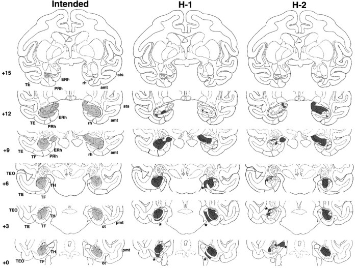
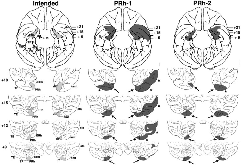
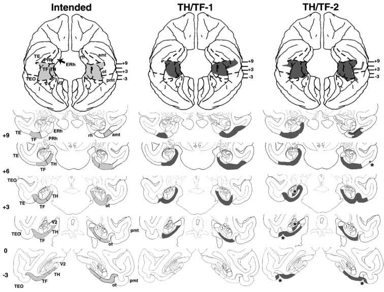
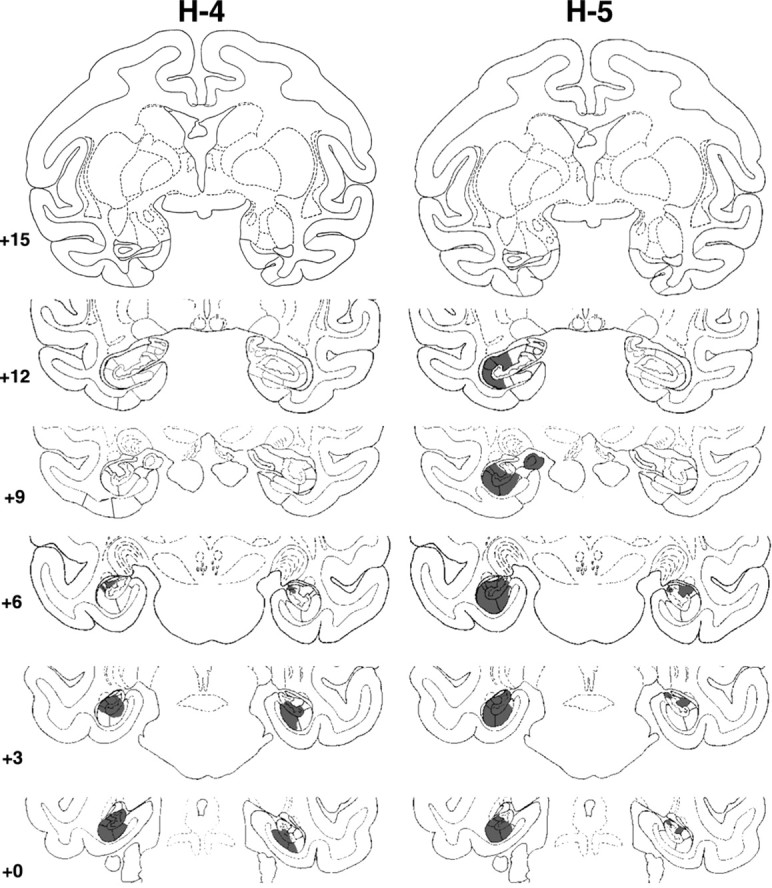
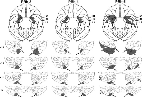
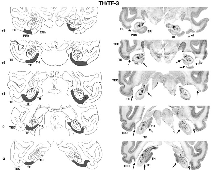
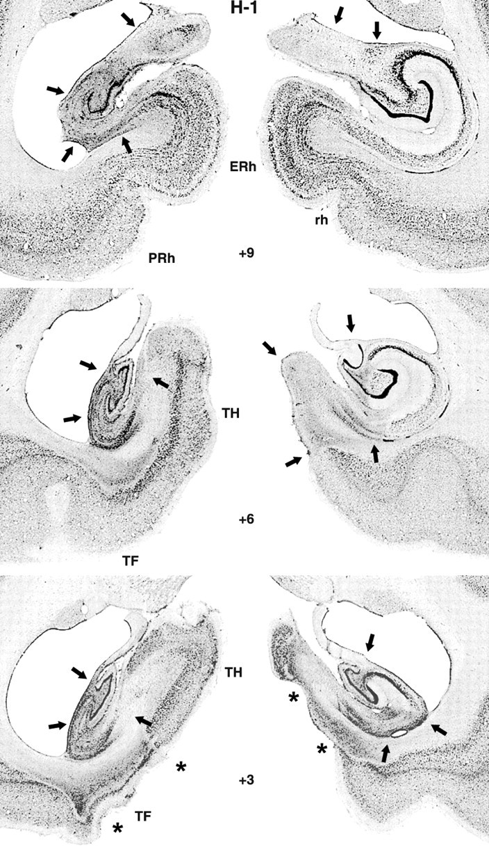
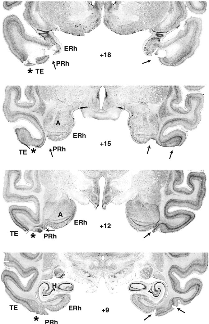
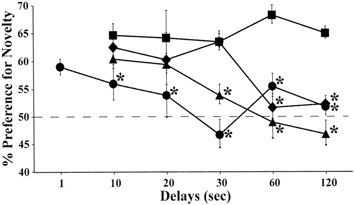
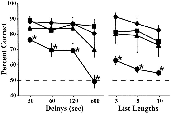
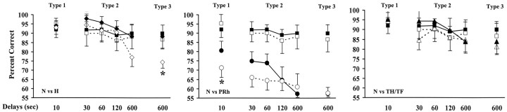
Recognition memory was assessed by submitting the same adult monkeys to visual paired comparison (VPC) with mixed delays (10-120 sec), followed by three consecutive versions of object-delayed nonmatching-to-sample (DNMS): increasing delays (10-600 sec), lengthened lists (3-10 objects), and intervening distractors in the delays (light at 10 sec, motor task at 30-600 sec, or context change at 600 sec). Four groups were tested: normal controls, monkeys with ibotenic acid lesions of the hippocampal formation (H), and monkeys with aspiration lesions of either the perirhinal (PRh) or parahippocampal (areas TH/TF) cortex. Group H was impaired on VPC at delays > or =60 sec but had difficulty on DNMS only at 600 sec delays with distraction. In group TH/TF, the VPC impairment emerged earlier (30 sec); yet, once the nonmatching rule was mastered, no significant change occurred on any DNMS condition. Only group PRh behaved congruently on VPC and DNMS, exhibiting a deficit at the easiest condition that worsened with increasing delays as well as in DNMS lengthened list and distraction conditions. These results led us to postulate that VPC and DNMS, as previously administered to monkeys, were not equivalent visual recognition memory probes. Specifically, we propose that, for VPC, because of passive (incidental) encoding, the animal's performance rests on both item familiarity and event recollection, whereas, for DNMS, because of active (purposeful) encoding, performance relies more on item familiarity. This proposal converges with current models postulating distinct, but interactive, mnemonic roles for the hippocampal and adjacent TH/TF regions.


Figures

Figure 1.
Figure 1.
Coronal sections through the hippocampal formation depicting the intended damage (left), shown in gray, and actual damage in cases H-1 and H-2 with neurotoxic lesions of the hippocampal formation, shown in black. The numerals on the left of each coronal section of the intended lesions indicate the distance in millimeters from the interaural plane. The asterisks point to unintended damage to adjacent structures. amt, Anterior middle temporal sulcus; ERh, entorhinal cortex; pmt, posterior middle temporal sulcus; rh, rhinal sulcus; ot, occipitotemporal sulcus; sts, superior temporal sulcus; TE, TEO, TH and TF, cytoarchitectonic fields described by von Bonin and Bailey (1947).
Figure 4.
Figure 4.
Coronal sections through the PRh cortex, depicting the intended damage (left), shown in gray, and actual damage in cases PRh-1 and PRh-2 with aspiration lesions of the PRh cortex, shown in black. The numerals on the left of each coronal section of the intended lesions indicate the distance in millimeters from the interaural plane. The asterisks point to unintended damage to adjacent structures. amt, Anterior middle temporal sulcus; ERh, entorhinal cortex; pmt, posterior middle temporal sulcus; rh, rhinal sulcus; ot, occipitotemporal sulcus; sts, superior temporal sulcus; TE, TEO, TH and TF, cytoarchitectonic fields described by von Bonin and Bailey (1947).
Figure 7.
Figure 7.
Coronal sections through areas TH and TF, depicting the intended damage (left), shown in gray, and actual damage in cases TH/TF-1 and TH/TF-2 with aspiration lesions of areas TH/TF, shown in black. The numerals on the left of each coronal section of the intended lesions indicate the distance in millimeters from the interaural plane. The asterisks point to unintended damage to adjacent structures. amt, Anterior middle temporal sulcus; ERh, entorhinal cortex; pmt, posterior middle temporal sulcus; rh, rhinal sulcus; ot, occipitotemporal sulcus; sts, superior temporal sulcus; TE, TEO, TH and TF, cytoarchitectonic fields described by von Bonin and Bailey (1947); V2, visual extrastriate cortical areas.
Figure 2.
Figure 2.
Coronal sections through the actual damage in cases H-4 and H-5 with neurotoxic lesions of the hippocampal formation, shown in black. The numerals on the left of each coronal section of the intended lesions indicate the distance in millimeters from the interaural plane. The asterisks point to unintended damage to adjacent structures.
Figure 5.
Figure 5.
Coronal sections through the actual damage in cases PRh-3, -4, and -5 with aspiration lesions of the PRh cortex, shown in black. The numerals on the left of each coronal section of the intended lesions indicate the distance in millimeters from the interaural plane. The asterisks point to unintended damage to adjacent structures.
Figure 8.
Figure 8.
Coronal sections through areas TH and TF (left) and corresponding photomicrographs of thionin-stained coronal sections (right) of case TH/TF-3. Note that fiber damage just below areas TH/TF was seen on histological sections but not on the postsurgical T1-weighed MR images. The numerals on the left of each coronal section of the intended lesions indicate the distance in millimeters from the interaural plane. The asterisks point to unintended damage to adjacent structures. ERh, Entorhinal cortex; pmt, posterior middle temporal sulcus; rh, rhinal sulcus; ot, occipitotemporal sulcus; sts, superior temporal sulcus; TE, TEO, TH and TF, cytoarchitectonic fields described by von Bonin and Bailey (1947).
Figure 3.
Figure 3.
Photomicrographs of thionine-stained coronal sections from a monkey with a hippocampal lesion (case H-1). The arrows indicate sites of cell death. Notice that the lesion extended through the entire left hippocampal formation, whereas it spared the lateralmost portion of the right hippocampal formation. The asterisks point to bilateral unintended damage to areas TH and TF (see level +3). ERh, Entorhinal cortex; rh, rhinal sulcus; TH and TF, cytoarchitectonic fields described by von Bonin and Bailey (1947).
Figure 6.
Figure 6.
Photomicrographs of thionin-stained coronal sections from a monkey with a PRh lesion (case PRh-4). ERh, Entorhinal cortex; TE, cytoarchitectonic fields described by von Bonin and Bailey(1947).
Figure 9.
Figure 9.
Average percentage of looking time at the novel stimulus during each delay condition of the VPC task for animals with hippocampal lesions (♦), animals with PRh lesions (•), animals with areas TH/TF lesions (▴), and unoperated controls (▪). The dashed horizontal line represents chance performance (50%), and vertical bars for each data point indicate SEM. *p < 0.05, compared with controls.
Figure 10.
Figure 10.
Average percentage of correct choices at the four delays (s-DNMS) and three lists (l-DNMS) of the DNMS task for each animal group. ♦, Animals with hippocampal lesions; •, animals with PRh lesions; ▴, animals with areas TH/TF lesions; ▪, unoperated controls. The dashed horizontal line represents chance performance (50%), and vertical bars for each data point indicate SEM. *p < 0.05, compared with controls.
Figure 11.
Figure 11.
Average percentage of correct choices in the standard (solid line) and distraction (dashed line) trials of the dDNMS for each distraction type in the four animal groups. ♦, Animals with hippocampal lesions; •, animals with PRh lesions; ▴, animals with areas TH/TF lesions; ▪, unoperated controls. The dashed horizontal line represents chance performance (50%), and vertical bars for each data point indicate SEM. *p < 0.05, compared with controls.

Similar articles

Cited by

References

    1. Alvarado MC, Bachevalier J (2003) Damage to perirhinal and parahippocampal TH/TF cortices in monkeys impairs performance on transverse patterning. Soc Neurosci Abstr 29: 324.3.
    1. Alvarado MC, Mishkin M, Bachevalier J (1998) Neurotoxic lesions of the hippocampal formation impair monkeys' acquisition of the transverse patterning. Soc Neurosci Abstr 24: 928.
    1. Bachevalier J, Nemanic S, Alvarado MC (2002) The medial temporal lobe structures and object recognition memory in nonhuman primates. In: Neuropsychology of memory, Ed 2 (Squire LR, Schacter DL, eds), pp 326-338. New York: Guilford.
    1. Baxter MG, Murray EA (2001) Opposite relationship of hippocampal and rhinal cortex damage to delayed nonmatching-to-sample deficits in monkeys. Hippocampus 11: 61-71. - PubMed
    1. Beason-Held LL, Rosene DL, Killiany RJ, Moss MB (1999) Hippocampal formation lesions produce memory impairment in the rhesus monkey. Hippocampus 9: 562-574. - PubMed

Publication types