

The long-term stability of new hippocampal place fields requires new protein synthesis
- PMID: 14985509
- PMCID: PMC373518
- DOI: 10.1073/pnas.0400385101
The long-term stability of new hippocampal place fields requires new protein synthesis
Abstract
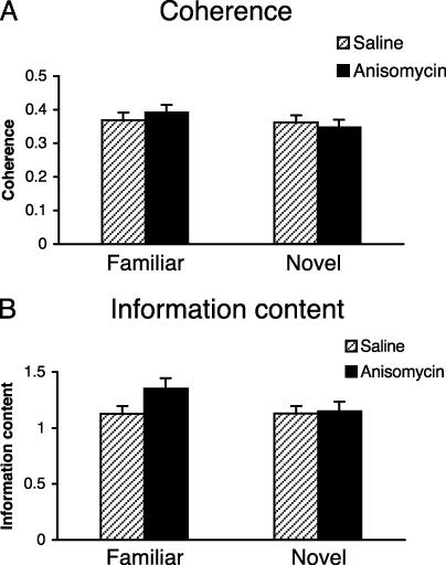
The hippocampus is critical for formation of spatial memories. Hippocampal pyramidal neurons in freely behaving animals exhibit spatially selective firing patterns, which taken together form an internal representation of the environment. This representation is thought to contribute to the hippocampal spatial memory system. Behavioral long-term memories differ from short-term memories in requiring the synthesis of new proteins. Does the development of the internal hippocampal representation also require the synthesis of new proteins? We found that blocking protein synthesis in the brain of mice by 95% does not affect short-term stability of newly formed hippocampal place fields but abolishes stability in the long term. By contrast, inhibiting protein synthesis does not affect the retention and recall of previously established fields in a familiar environment, indicating that protein synthesis-dependent reconsolidation is not required for recall. Our results indicate that place fields parallel both behavioral memories and the late phase of long-term potentiation in requiring the synthesis of new proteins for consolidation.
Figures


Similar articles
-
Protein degradation, as with protein synthesis, is required during not only long-term spatial memory consolidation but also reconsolidation.Eur J Neurosci. 2008 Jun;27(11):3009-19. doi: 10.1111/j.1460-9568.2008.06262.x. Eur J Neurosci. 2008. PMID: 18588539
-
Inhibition of hippocampal protein synthesis following recall disrupts expression of episodic-like memory in trace conditioning.Hippocampus. 2005;15(3):333-9. doi: 10.1002/hipo.20055. Hippocampus. 2005. PMID: 15523611
-
Long-term memory underlying hippocampus-dependent social recognition in mice.Hippocampus. 2000;10(1):47-56. doi: 10.1002/(SICI)1098-1063(2000)10:1<47::AID-HIPO5>3.0.CO;2-6. Hippocampus. 2000. PMID: 10706216
-
Molecular, cellular, and neuroanatomical substrates of place learning.Neurobiol Learn Mem. 1998 Jul-Sep;70(1-2):44-61. doi: 10.1006/nlme.1998.3837. Neurobiol Learn Mem. 1998. PMID: 9753586 Review.
-
Hippocampal place cells, context, and episodic memory.Hippocampus. 2006;16(9):716-29. doi: 10.1002/hipo.20208. Hippocampus. 2006. PMID: 16897724 Review.
Cited by
-
A stable hippocampal representation of a space requires its direct experience.Proc Natl Acad Sci U S A. 2011 Aug 30;108(35):14654-8. doi: 10.1073/pnas.1105445108. Epub 2011 Aug 18. Proc Natl Acad Sci U S A. 2011. PMID: 21852575 Free PMC article.
-
Altered Hippocampal Place Cell Representation and Theta Rhythmicity following Moderate Prenatal Alcohol Exposure.Curr Biol. 2020 Sep 21;30(18):3556-3569.e5. doi: 10.1016/j.cub.2020.06.077. Epub 2020 Jul 23. Curr Biol. 2020. PMID: 32707066 Free PMC article.
-
Extinction of Learned Fear Induces Hippocampal Place Cell Remapping.J Neurosci. 2015 Jun 17;35(24):9122-36. doi: 10.1523/JNEUROSCI.4477-14.2015. J Neurosci. 2015. PMID: 26085635 Free PMC article.
-
Hippocampal Place Fields Maintain a Coherent and Flexible Map across Long Timescales.Curr Biol. 2018 Nov 19;28(22):3578-3588.e6. doi: 10.1016/j.cub.2018.09.037. Epub 2018 Nov 1. Curr Biol. 2018. PMID: 30393037 Free PMC article.
-
Abnormal c-Fos expression in TetTag mice containing fos-EGFP.Front Behav Neurosci. 2024 Dec 17;18:1500794. doi: 10.3389/fnbeh.2024.1500794. eCollection 2024. Front Behav Neurosci. 2024. PMID: 39741565 Free PMC article.
References
-
- Squire, L. R. & Kandel, E. R. (1999) Memory: From Mind To Molecules (Scientific American Library, New York).
-
- O'Keefe, J & Nadel, L. (1978) The Hippocampus as a Cognitive Map (Clarendon Press, Oxford, U.K.).
Publication types
MeSH terms
Substances
LinkOut - more resources
Full Text Sources
Medical
