Review
. 2004 May;13(3):184-92.
doi: 10.1007/s00586-003-0654-4. Epub 2004 Feb 25.

Kyphoplasty for treatment of osteoporotic vertebral fractures

Affiliations
Review

Kyphoplasty for treatment of osteoporotic vertebral fractures

Paul F Heini et al. Eur Spine J. 2004 May.

Abstract

Cement reinforcement for the treatment of osteoporotic vertebral fractures is efficient mean with high success in pain release and prevention of further sintering of the reinforced vertebrae; however, the technique does not allow to address the kyphotic deformity. Kyphoplasty was designed to address the kyphotic deformity and help to realign the spine. It involves the percutaneous placement of an inflatable bone tamp into a vertebral body. Restoration of VB height and kyphosis correction is achieved by inflation of the bone tamp with liquid. After deflation, a cavity is created that eases the cement application. The potential of kyphosis reduction is given in fresh fractures with a range of 0-90% for height restoration and absolute correction of the kyphotic angle of 8.5 degrees. The cavity formation, on one hand, and the different cementing technique leads to lower risk for cement extravasation. An alternative method for kyphosis correction represents the so-called lordoplasty where the adjacent vertebrae are reinforced first and with the cannulas in place acting as a lever the reduction of the collapsed vertebra can be performed. The results with respect to kyphosis correction are superior in comparison with a kyphoplasty procedure.


Figures

Fig. 1
Fig. 1
The pedicles serving as landmark. A K-wire is inserted under biplanar fluoroscopic control. Ideally, the final position of the balloons should be centered between the end plates within the anterior two-thirds of the vertebral body (see Acknowledgements)
Fig. 2
Fig. 2
Five steps of kyphoplasty: 1 placement of a guide wire; 2 insertion of a working cannulas; 3 reaming of working channels; 4 inflation of the balloons, and 5 injection of polymethylmetacrylate (see Acknowledgements)
Fig. 3a, b
Fig. 3a, b
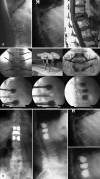
A 68-year-old woman. a Pincer-type fracture of L2 after minor trauma. In order to restore the end-plate impression, a combined procedure with internal fixation and bisegmental fusion and balloon kyphoplasty was planned. b Intraoperative sequence with internal fixation in place and inflation of the balloons. The end plates have been pushed in a better position with the balloons. After deflation, the void in the vertebral body is well visible; however, the reduction of the end plate is lost, to a certain degree. c Standing X-ray after the procedure and follow-up control after 1 year. There is some local cement leakage on the left side which is clinically not symptomatic. After 1 year, the alignment of the spine is preserved. There is spontaneous ossification at anterior longitudinal ligament. The patient is doing well
Fig. 4a–c
Fig. 4a–c
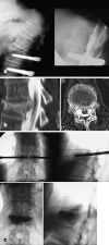
This 73-year-old woman was fused from L3 to S1 in former years and was surgically treated for a spinal stenosis at L2–L3 and stabilized with a semi-rigid fixation system. From the beginning, after the surgery, the patient complaint about sharp, localized back pain and became bedridden. a The initial X-ray shows a simple compression fracture of T12. After 2 months, an important kyphotic deformity developed with spinal canal narrowing. The patient was foreseen for an open combined anterior and posterior procedure by her treating spine surgeon; however, due to the bad general health condition, the anesthesiologist considered such a procedure not appropriate. b The CT reconstruction shows a certain spontaneous correction of the deformity in the supine position, which in turn is a hint of an instability; therefore, a kyphoplasty procedure was performed under general anesthesia. c In the prone position there is already a marked improvement of the kyphosis. It was possible to create the working channels perfectly, centered in the vertebral body. The kyphoplasty procedure did restore the kyphotic deformity further, and the situation after the cementing shows a well-established alignment of the spine. The patient regained his former activity level after 1 week
Fig. 5a, b
Fig. 5a, b
Potential problems related to cement reinforcement. A balloon kyphoplasty was performed in this 80-year-old patient. a The postoperative standing film shows an accurate cement filling with a regular alignment of the spine. After 2 months, the patient presents with a new fracture of the adjacent vertebra. There is some dislodgement of the cement plug. The standing X-ray shows the segmental collapse T12–L1, and the image taken with the patient lying supine depicts a major defect and obvious instability at T12–L1. b Because of the bad general condition, the solution in this case was a percutaneous filling of this defect and a reinforcement of the adjacent vetebrae. The patient was put under general anesthesia and placed in hyperextension. The standing X-ray after 6 months shows a well-preserved alignment. The patient is doing well. She does complain about lower back pain that is not related to the changes at thoraco-lumbar junction
Fig. 6
Fig. 6
Loss of reduction after balloon deflation and cementing indicates an inherent problem related to the technique of kyphoplasty. The vertebral body height was restored to 85% normal with the balloons inflated. After cementing, the resulting correction seen on the standing X-ray is only half of the intraoperative situation
Fig. 7a–c
Fig. 7a–c
Natural history of osteoporotic fractures in a 73-year-old woman. a Both fractures (T11 and L2) were qualified as old as the patient had only minor complaints. Over a period of 4 months, the patient complained about increasing pain. The standing X-ray shows the severe kyphotic deformity at T11. The MRI scan shows a nearly complete collapse of T11, but furthermore, some degree of spontaneous correction of the kyphosis. b Alternative method of kyphosis correction: lordoplasty means cementing of the adjacent vertebrae in a classical manner. When the cement is dried, the cannulas are used as a lever in order to apply a lordotic moment. The facet joints are acting as a hypomochlion. The collapsed vertebral body has gained height (radiolucent area), and the kyphotic deformity is corrected to a major extent. Cementing of the fractured vertebrae: when the cement is cured, the tension is released and the cannulas are removed. c Standing X-ray, 6 months postoperatively. The patient is free of pain in unrestricted activity

Comment in

References

    1. Barr Spine. 2000;25:923. - PubMed
    1. Belkoff Spine. 2002;27:1640. doi: 10.1097/00007632-200208010-00009. - DOI - PubMed
    1. Belkoff Am J Neuroradiol. 2001;22:1212. - PMC - PubMed
    1. Belkoff Spine. 2001;26:151. doi: 10.1097/00007632-200101150-00008. - DOI - PubMed
    1. Berlemann J Bone Joint Surg Br. 2002;84:748. - PubMed