. 2004 Mar 24;23(6):1279-88.
doi: 10.1038/sj.emboj.7600137. Epub 2004 Feb 26.

Bsd2 binds the ubiquitin ligase Rsp5 and mediates the ubiquitination of transmembrane proteins

Affiliations

Bsd2 binds the ubiquitin ligase Rsp5 and mediates the ubiquitination of transmembrane proteins

Ewald H Hettema et al. EMBO J. .

Abstract

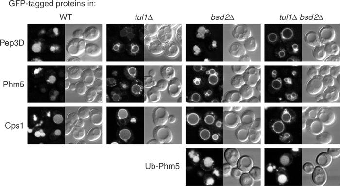
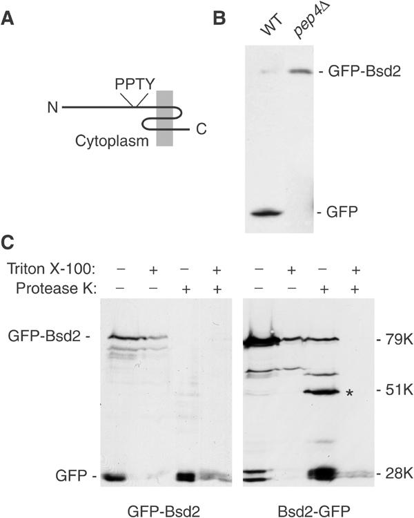
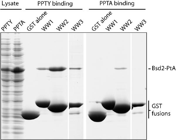
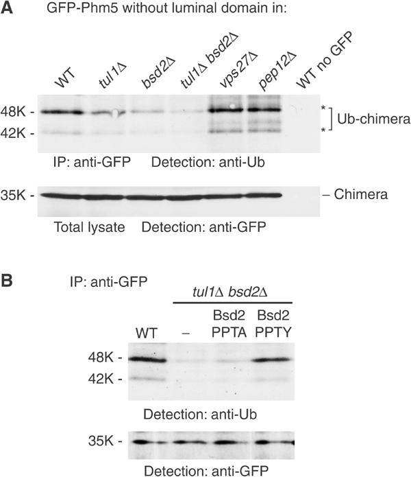
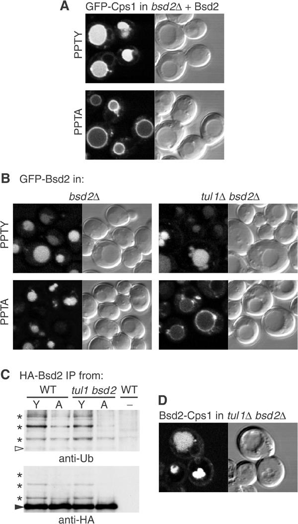
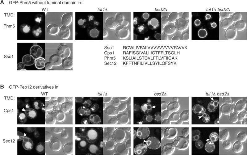
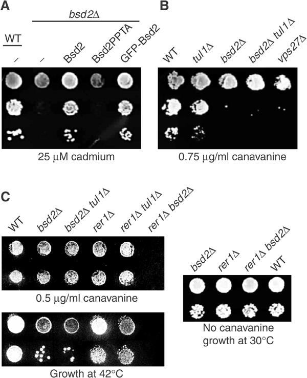
Membrane proteins destined for the vacuolar or lysosomal lumen are typically ubiquitinated, the ubiquitin serving as a targeting signal for the multivesicular body pathway. The RING-domain ubiquitin ligase Tul1 is an integral membrane protein that modifies the yeast vacuolar enzyme carboxypeptidase S (Cps1), the polyphosphatase Ppn1/Phm5 and other proteins containing exposed hydrophilic residues within their transmembrane domains (TMDs). Here we show that Bsd2 provides an alternative ubiquitination mechanism for Cps1, Phm5 and other proteins. Bsd2 is a three-TMD protein with a PPXY motif that binds the HECT domain ubiquitin ligase Rsp5. It can thus act as a specific adaptor linking Rsp5 to its substrates. Like Tul1, the Bsd2 system recognises polar TMDs. Bsd2 also controls the vacuolar targeting of a manganese transporter and a mutant plasma membrane ATPase, and together with the ER retrieval receptor Rer1, it protects cells from stress. We suggest that Bsd2 has a wide role in the quality control of membrane proteins. Bsd2 is the yeast homologue of human NEDD4 binding protein N4WBP5, which may therefore have similar functions.


Figures

Figure 1
Figure 1
Bsd2 contributes to the sorting of proteins into MVBs. GFP-tagged proteins were expressed in the indicated strains and examined by confocal microscopy. Representative examples of the GFP fluorescence and the corresponding interference contrast images are shown.
Figure 2
Figure 2
Orientation and fate of Bsd2. (A) Predicted structure of Bsd2, with the position of the PPTY motif indicated. The protein is 321 residues long. (B) Immunoblotting with anti-GFP of an N-terminal GFP fusion protein expressed from its own promoter in bsd2Δ cells that were wild type for protease activity (WT), and in a strain deficient in vacuolar proteases (pep4Δ). (C) Membranes from cells expressing Bsd2 tagged with GFP at the N terminus (GFP-Bsd2) or the C terminus (Bsd2-GFP) were digested with protease K with or without 1% Triton X-100 as indicated, and the fragments were detected by immunoblotting. Numbers indicate the apparent sizes of the proteins relative to globular protein markers. Note that Bsd2 and its fragments appear larger than they really are. The asterisk indicates a prominent proteolytic fragment with an apparent size of 51 kDa.
Figure 3
Figure 3
Binding of the Bsd2 N terminus to the WW domains of Rsp5. A Coomassie-stained gel is shown. E. coli lysates (left lanes) containing either the PPTY or PPTA versions of the Bsd2 N terminus fused to protein A (Bsd2-PtA) were incubated with beads containing GST alone or fused to one of the three WW domains of Rsp5. After extensive washing, the GST fusions and bound proteins were eluted with SDS sample buffer and subjected to gel electrophoresis.
Figure 4
Figure 4
Bsd2 affects ubiquitination of Phm5. (A) A GFP-tagged variant of Phm5 lacking the luminal domain was immunoprecipitated from the indicated strains, a strain not expressing the GFP-tagged protein serving as a control. Ubiquitinated species were then identified by immunoblotting with anti-ubiquitin. Asterisks indicate the two major ubiquitinated species. These species could also be detected, although inefficiently, by blotting with anti-GFP (not shown). Samples of the total cell lysates were run in parallel and immunoblotted to show the equivalent amounts of intact GFP-Phm5 chimera in each sample. (B) As (A) except that tul1 bsd2 double mutant cells contained either an empty vector plasmid (−), or plasmids expressing untagged wild-type (PPTY) or point mutant (PPTA) Bsd2 from its own promoter. Samples of the immunoprecipitates were also blotted with anti-GFP to show that they contained equivalent amounts of GFP-Phm5.
Figure 5
Figure 5
Effects of the PPTY motif on sorting of Cps1 and Bsd2. (A) Complementation of a bsd2 null mutant by expression of untagged wild-type (PPTY) Bsd2 restores sorting of Cps1 into vacuoles, whereas the point mutant (PPTA) does not. (B) Expression of GFP-Bsd2 with wild-type (PPTY) or mutant (PPTA) sequences in the indicated strains. Note that entry into vacuoles requires either the PPTY sequence or Tul1. (C) HA-tagged Bsd2 containing the normal (Y) or mutated (A) PPTY motif was immunoprecipitated from wild-type or tul1 bsd2 cells and probed with antiubiquitin; the blot was then re-probed with anti-HA. The arrowhead indicates the position of full-length HA-Bsd2 (apparent size 54 kDa), and the asterisks indicate ubiquitinated forms. Control cells (−) expressed no HA-tagged protein. (D) A chimera consisting of the N-terminal cytoplasmic domain of Bsd2 fused to GFP-Cps1 is sorted into vacuoles in a tul1 bsd2 double mutant.
Figure 6
Figure 6
Sorting depends on TMDs. (A) GFP-Phm5 constructs in which the entire luminal domain was deleted were expressed in the indicated strains. The constructs had either the normal Phm5 TMD or the Sso1 TMD as indicated. The sequences of these, and the Cps1 and Sec12 TMDs for comparison, are shown. (B) GFP-Pep12 constructs in which the Pep12 TMD had been replaced with that from Cps1 or Sec12 were expressed in the indicated strains. Note that Pep12 itself is never found inside vacuoles, and thus entry of these constructs is dictated entirely by their TMDs. Arrows indicate nuclear envelope and peripheral ER.
Figure 7
Figure 7
Stress sensitivity of bsd2 mutants. (A) Serial dilutions of cells were grown on plates containing 25 μM cadmium chloride. The bsd2Δ cells contained either an empty vector plasmid (−), or one expressing untagged wild-type Bsd2 or the PPTA mutant (Bsd2PPTA) from the BSD2 promoter, or a plasmid expressing GFP-Bsd2. (B) Serial dilutions of strains with the indicated genotypes were grown on plates lacking arginine and containing 0.75 μg/ml canavanine. Note that growth strongly depends on the number of cells plated, since uptake of canavanine by the cells depletes it from the plate. All strains were plated at the same densities. (C) Cells were grown on plates containing 0.5 μg/ml canavanine, or on normal plates that were incubated either at 30 or 42°C.

References

    1. Beck T, Schmidt A, Hall MN (1999) Starvation induces vacuolar targeting and degradation of the tryptophan permease in yeast. J Cell Biol 146: 1227–1238 - PMC - PubMed
    1. Bilodeau PS, Urbanowski JL, Winistorfer SC, Piper RC (2002) The Vps27p Hse1p complex binds ubiquitin and mediates endosomal protein sorting. Nat Cell Biol 4: 534–539 - PubMed
    1. Black MW, Pelham HR (2000) A selective transport route from Golgi to late endosomes that requires the yeast GGA proteins. J Cell Biol 151: 587–600 - PMC - PubMed
    1. Cristillo AD, Nie L, Macri MJ, Bierer BE (2003) Cloning and characterization of N4WBP5A, an inducible, cyclosporine-sensitive, Nedd4-binding protein in human T lymphocytes. J Biol Chem 278: 34587–34597 - PubMed
    1. Dupre S, Haguenauer-Tsapis R (2001) Deubiquitination step in the endocytic pathway of yeast plasma membrane proteins: crucial role of Doa4p ubiquitin isopeptidase. Mol Cell Biol 21: 4482–4494 - PMC - PubMed

Publication types

MeSH terms