Case Reports
. 2004 Apr;74(4):731-7.
doi: 10.1086/383094. Epub 2004 Feb 25.

A dual phenotype of periventricular nodular heterotopia and frontometaphyseal dysplasia in one patient caused by a single FLNA mutation leading to two functionally different aberrant transcripts

Affiliations
Case Reports

A dual phenotype of periventricular nodular heterotopia and frontometaphyseal dysplasia in one patient caused by a single FLNA mutation leading to two functionally different aberrant transcripts

Martin Zenker et al. Am J Hum Genet. 2004 Apr.

Abstract

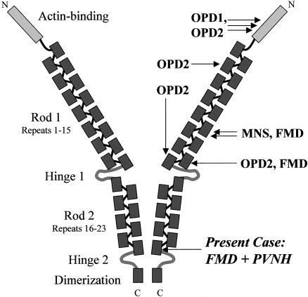
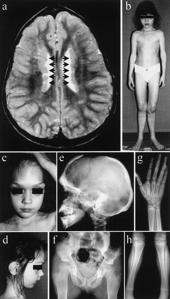
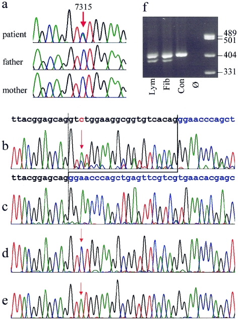
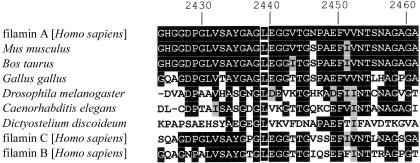
Two disorders, periventricular nodular heterotopia (PVNH) and a group of skeletal dysplasias belonging to the oto-palato-digital (OPD) spectrum, are caused by FLNA mutations. They are considered mutually exclusive because of the different presumed effects of the respective FLNA gene mutations, leading to loss of function (PVNH) and gain of function (OPD), respectively. We describe here the first patient manifesting PVNH in combination with frontometaphyseal dysplasia, a skeletal dysplasia of the OPD-spectrum. A novel de novo mutation, 7315C-->A in exon 45 of the FLNA gene, was identified. It leads to two aberrant transcripts, one full-length transcript with the point mutation causing a substitution of a highly conserved leucine residue (L2439M) and a second shortened transcript lacking 21 bp due to the creation of an ectopic splice donor site in exon 45. We propose that the dual phenotype is caused by two functionally different, aberrant filamin A proteins and therefore represents an exceptional model case of allelic gain-of-function and loss-of-function phenotypes due to a single mutational event.


Figures

Figure  1
Figure 1
Diagram showing the structure of the filamin A homodimer, functional domains (left), and localization of the previously described and the novel mutations (right). Adapted from Stossel et al. (2001).
Figure  2
Figure 2
a, Cranial MRI of the patient at age 5 years, showing typical PVNHs (arrows). b–d, Clinical photographs of the patient at age 7 years: massively prominent supraorbital ridges; large-appearing eyes with hypertelorism; receding, pointed chin; slender, deformed thorax; genua valga et recurvata; and extension deficit in the elbows. e–h, Radiological findings of the patient at age 7 years. e, Skull radiograph showing increased density of the skull, with deficient pneumatization of mastoid and sinuses and a widely open anterior fontanel. f, Radiograph of the pelvis and hips, showing coxa valga et antetorta, with widened femoral necks. Radiographs of the right distal forearm and hand (g) and of the knees and lower legs (h), showing metaphyseal widening of long bones, short terminal phalanges, genua valga, and slight bending of tibiae and fibulae.
Figure  3
Figure 3
a, Sequence traces from genomic DNA of the patient and her clinically normal parents, showing the heterozygous C→A exchange position, 7315, in the patient and absence of the mutation in both parents. b–e, Demonstration of the expression of two different aberrant FLNA transcripts in fibroblasts. b, Electropherogram derived from sequencing of the RT-PCR products from fibroblast mRNA, showing the presence of a shortened alternative transcript lacking 21 bases from exon 45. The normal sequence of the exons 45 and 46 are indicated above in black and blue, respectively. The position of the point mutation 7315C→A is depicted in red and marked by red arrows. Note that the T peak at position of the mutation is higher than the C representing the wild-type allele. Predominance of the mutant allele reflects the X-inactvation pattern. c–e, Sequences from different clones of the RT-PCR product cloned in E. coli. All three transcripts predicted from the sequencing results in panel b could be detected: 21-bp deletion (c), wild-type allele (d), and point mutation (e). f, Electrophoresis of a 409-bp RT-PCR fragment on a 2.5% agarose gel, showing presence of a shortened fragment in the patient's lymphocytes (lym) and fibroblasts (fib) but not in a normal male control (con). Ø = no template control.
Figure  4
Figure 4
Three-dimensional models of the filamin A rod domain repeat unit 23, generated on the basis of the homologous structures of the 4th (Fucini et al. 1997) and 5th (McCoy et al. 1999) module of the F-Actin Cross-Linking Gelation Factor (Abp-120) by use of Swiss-Model (Guex and Peitsch 1997; Protein Data Bank). a, Ribbon model of a repeat unit showing, in cyan, the regular position of the strand deleted in the shortened protein. Amino- and carboxyterminal ends of the repeat are marked by N- and -C, respectively. b, Model showing the respective position of the leucine residue corresponding to position 2439 depicted in cyan. It resides in a hydrophobic surface cluster. Nonpolar residues are shown in red.
Figure  5
Figure 5
Alignment of the beginning of filamin A repeat 23 and filamin A homologues from various species, as well as the human paralogues filamin B and C (GenBank), showing conservation of the leucine residue at position 2439 (white frames on black background). The conserved amino acids are shown with black background and similar amino acids with a gray background. Numbers above indicate amino acid position in the filamin A protein (GenBank accession number NP_001447).

Similar articles

Cited by

References

Electronic-Database Information

    1. dbSNP Home Page, http://www.ncbi.nih.gov/SNP/ (for IVS45-64T→C [accession number rs2070819] and coding SNPs within FLNA [accession number rs2070825] and TIMP1 [accession number rs4898])
    1. GenBank, http://www.ncbi.nih.gov/Genbank/ (for FLNA [accession numbers: genomic: NT_025965; mRNA: NM_001456; protein: NP_001447; GenBank: X53416], FLNA protein [Mus musculus: gi|13278531|gb|AAH04061.1], filamin A [Bos taurus: gi|18377580|gb|AAL66773.1], for filamin A [Gallus gallus: gi|15341202|dbj|BAB63943.1], filamin C [Homo sapiens: gi|4218955|gb|AAD12245.1], filamin B [H. sapiens: gi|3298597|gb|AAC39842.1], filamin1 [Drosophila melanogaster: gi|6707288|gb|AAF25614.1|AF174492_1], Filamin/ABP280 repeat family member (4B112) [Caenorhabditis elegans: gi|17543918|ref|NP_499911.1])
    1. Human Gene Mutation Database, http://archive.uwcm.ac.uk/uwcm/mg/hgmd0.html (for known FLNA mutations in PVNH)
    1. Online Mendelian Inheritance in Man (OMIM), http://www.ncbi.nlm.nih.gov/Omim/ (for PVNH, OPD1, OPD2, MNS, and FMD)
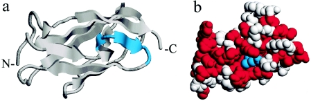
    1. Protein Data Bank, http://www.rcsb.org/pdb/ (for the structures of the 4th [1KSR] and 5th [1QFH] module of the F-actin cross-linking gelation factor [Abp-120])

References

    1. Allen RC, Zoghbi HY, Moseley AB, Rosenblatt HM, Belmont JW (1992) Methylation of HpaII and HhaI sites near the polymorphic CAG repeat in the human androgen-receptor gene correlates with X chromosome inactivation. Am J Hum Genet 51:1229–1239 - PMC - PubMed
    1. Carrel L, Willard HF (1996) An assay for X inactivation based on differential methylation at the fragile X locus, FMR1. Am J Med Genet 64:27–3010.1002/(SICI)1096-8628(19960712)64:1<27::AID-AJMG3>3.0.CO;2-O - DOI - PubMed
    1. Cartegni L, Chew SL, Krainer AR (2002) Listening to silence and understanding nonsense: exonic mutations that affect splicing. Nat Rev Genet 3:285–29810.1038/nrg775 - DOI - PubMed
    1. Fox JW, Lamperti ED, Eksioglu YZ, Hong SE, Feng Y, Graham DA, Scheffer IE, Dobyns WB, Hirsch BA, Radtke RA, Berkovic SF, Huttenlocher PR, Walsh CA (1998) Mutations in filamin 1 prevent migration of cerebral cortical neurons in human periventricular heterotopia. Neuron 21:1315–1325 - PubMed
    1. Fucini P, Renner C, Herberhold C, Noegel AA, Holak TA (1997) The repeating segments of the F-actin cross-linking gelation factor (ABP-120) have an immunoglobulin-like fold. Nat Struct Biol 4:223–230 - PubMed

Publication types

MeSH terms