. 2004 Mar;86(3):1332-44.
doi: 10.1016/S0006-3495(04)74205-8.

Insights into the molecular mechanism of rotation in the Fo sector of ATP synthase

Affiliations

Insights into the molecular mechanism of rotation in the Fo sector of ATP synthase

Aleksij Aksimentiev et al. Biophys J. 2004 Mar.

Abstract

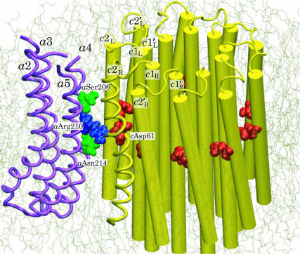
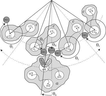
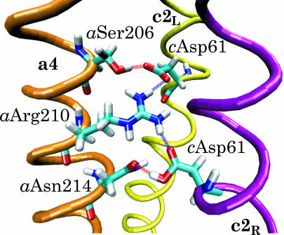
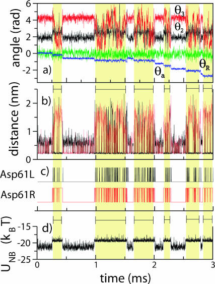
F(1)F(o)-ATP synthase is a ubiquitous membrane protein complex that efficiently converts a cell's transmembrane proton gradient into chemical energy stored as ATP. The protein is made of two molecular motors, F(o) and F(1), which are coupled by a central stalk. The membrane unit, F(o), converts the transmembrane electrochemical potential into mechanical rotation of a rotor in F(o) and the physically connected central stalk. Based on available data of individual components, we have built an all-atom model of F(o) and investigated through molecular dynamics simulations and mathematical modeling the mechanism of torque generation in F(o). The mechanism that emerged generates the torque at the interface of the a- and c-subunits of F(o) through side groups aSer-206, aArg-210, and aAsn-214 of the a-subunit and side groups cAsp-61 of the c-subunits. The mechanism couples protonation/deprotonation of two cAsp-61 side groups, juxtaposed to the a-subunit at any moment in time, to rotations of individual c-subunit helices as well as rotation of the entire c-subunit. The aArg-210 side group orients the cAsp-61 side groups and, thereby, establishes proton transfer via aSer-206 and aAsn-214 to proton half-channels, while preventing direct proton transfer between the half-channels. A mathematical model proves the feasibility of torque generation by the stated mechanism against loads typical during ATP synthesis; the essential model characteristics, e.g., helix and subunit rotation and associated friction constants, have been tested and furnished by steered molecular dynamics simulations.


Figures

FIGURE 1
FIGURE 1
Schematic view of the E. coli ATP synthase. The solvent-exposed F1 unit (top) consists of subunits α3β3γδɛ; the membrane Fo unit (bottom) consists of subunits ab2c10.
FIGURE 2
FIGURE 2
Microscopic model of Fo ATPase composed of a four-helix bundle (a2–a5) of subunit a and an oligomer of 10 c-subunits. Only backbones of subunit a and of one out of 10 cTMH-2 (c2R) are shown as tubes; the rest of the c10 oligomer's helices are shown as cylinders. The binding sites (cAsp-61) of the c10 oligomer, the termini of the proton half-channels (aSer-204 and aAsn-214), and the critical aArg-210 residues of subunit a are drawn in van der Waals representation. The interface between a- and c-subunits was modeled.
FIGURE 3
FIGURE 3
RMSD values of the a1c10 complex α-carbon atoms during the equilibration.
FIGURE 4
FIGURE 4
Stochastic model for Fo (view from cytoplasm). Four out of 10 c-subunits and the a-subunit are shown. The c10 complex is fixed, and the a-subunit can move in either direction (angle θa). This is equivalent to the more natural choice of a fixed subunit a and a moving c10 complex. The second transmembrane helix (c2) of each c-subunit can rotate independently (described by angles θ1, θ2, θ3, and θ4), thereby moving the key cAsp-61 residues, which are the proton-binding sites. The c1 helices do not rotate. Similarly, only the fourth helix of the a-subunit (a4) can rotate (angle θR), moving the aArg-210 residue; helices a2, a3, and a5 do not rotate. Proton transfer occurs between the terminal residue of the periplasmic channel (aAsn-214) and the cAsp-61 binding site on helix c2R, and between the terminal residue of the cytoplasmic channel (aSer-206) and the cAsp-61 binding site on c2L. Motions are confined to the plane of the figure. The system is fully described by helix orientations θ1, θ2, θ3, and θ4 (c-subunits), θR (a4), rotor angle θa, and protonation state of the two aspartates (cAsp-61) on helices c2L and c2R.
FIGURE 5
FIGURE 5
Potentials of mean force used in the stochastic simulations: a double-well potential governing rotation of the c2 helices (open squares) and a parabolic potential governing rotation of the a4 helix (open circles).
FIGURE 6
FIGURE 6
Schematic representation of the sequence of events suggested by our study. These events, labeled af, occur during rotation of the c10 oligomer by 2π/10 in the synthesis direction, viewed here from the cytoplasm. (a) In the starting conformation, two residues cAsp-61 are deprotonated and form a bidentate salt bridge with aArg-210, cAsp-61aArg-210–cAsp-61. (b) A proton is transferred from the terminal residue of the periplasmic proton channel, aAsn-214, to cAsp-61 on helix c2R. (c) Subunit a rotates clockwise with respect to the c10 oligomer in concert with a clockwise rotation of helix c2L. When subunit a approaches helix c2L′, cAsp-61 on that helix rotates by 180°. The latter rotation may proceed in either clockwise or counterclockwise direction. (d) The concerted rotation of subunit a and helix c2L are completed: cAsp-61 on helix c2L′ has rotated by 180° toward subunit a. (e) A proton is transferred to the terminal residue of the cytoplasmic proton channel, aSer-206. (f) The system returns to the starting conformation a, but with the c10 oligomer advanced by an angle 2π/10. We note that the processes depicted are of stochastic nature, and, hence, do not necessarily obey the strict sequence shown.
FIGURE 7
FIGURE 7
Hydrogen-bond network formed between the binding sites (cAsp-61) and the terminal residues of the proton periplasm (aAsn-214) and cytoplasm (aSer-206) channels. The critical residue aArg-210 forms transient hydrogen bonds with both binding sites.
FIGURE 8
FIGURE 8
Concerted rotation of the c-subunit outer helix and the c10 complex in a lipid bilayer. The c2L helix has been forced to rotate clockwise by 180°. Shown in the instance when the salt bridge is transferred between two neighboring c-subunits, i.e., when the conformation cAsp-61aArg-210–cAsp-61 has been momentarily assumed.
FIGURE 9
FIGURE 9
Stochastic events involved in Fo function. (a) Time evolution of helix angles θ2 (black), θ3 (red), θR (green), and rotor angle θa (blue). The angles are defined in Fig. 4. The a-subunit rotation takes place in discrete steps (blue line). (b) Distances between residues aArg-210–cAsp-61 of c2L (black) and aArg-210–cAsp-61 of c2R (red). Respective salt bridges are formed when these distances decrease below ∼0.25 nm. When both aspartates are deprotonated, a two-color pattern of lines at 0.25 nm indicates a frequent transfer of the salt bridge from one aspartate to another. When one of the aspartates is protonated (highlighted regions), a two-color pattern does not indicate a salt bridge transfer, but originates from the cyclic boundary conditions invoked when subunit a passes the boundary. (c) Protonation states of cAsp-61L (black) and cAsp-61R (red) (see text). (d) Nonbonded interaction energy of the three residues. The steps of the energy function are correlated with protonation/deprotonation of two cAsp-61 residues and the step motion of the a-subunit.
FIGURE 10
FIGURE 10
Substeps of a-subunit rotation. (Left) When both cAsp-61 residues are deprotonated, the average internal potential acting on the a-subunit is symmetric (dashed line). The F1 load (dotted line) shifts the minimum of the average potential to the right (solid line). This figure corresponds to steps a and f in Fig. 6. (Right) When cAsp-61 on c2R receives a proton from the periplasm, the average internal potential becomes asymmetric. The minimum of the total potential is shifted to the left in this case. This figure corresponds to steps c and d in Fig. 6.

References

    1. Abrahams, J., A. Leslie, R. Lutter, and J. Walker. 1994. Structure at 2.8-Å resolution of F1-ATPase from bovine heart mitochondria. Nature. 370:621–628. - PubMed
    1. Angevine, C. M., and R. H. Fillingame. 2003. Aqueous access channels in subunit a of rotary ATP synthase. J. Biol. Chem. 278:6066–6074. - PubMed
    1. Batcho, P. F., D. A. Case, and T. Schlick. 2001. Optimized particle-mesh Ewald/multiple-time step integration for molecular dynamics simulations. J. Chem. Phys. 115:4003–4018.
    1. Berg, H. editor. 1983. Random Walks in Biology. Princeton University Press, Princeton, NJ.
    1. Böckmann, R. A., and H. Grubmüller. 2002. Nanoseconds molecular dynamics simulation of primary mechanical energy transfer steps in F1-ATP synthase. Nat. Struct. Biol. 9:198–202. - PubMed

Publication types

Substances

LinkOut - more resources