. 2004 Mar;78(6):2729-37.
doi: 10.1128/jvi.78.6.2729-2737.2004.

Human rhinovirus type 2-antibody complexes enter and infect cells via Fc-gamma receptor IIB1

Affiliations

Human rhinovirus type 2-antibody complexes enter and infect cells via Fc-gamma receptor IIB1

Günther Baravalle et al. J Virol. 2004 Mar.

Abstract

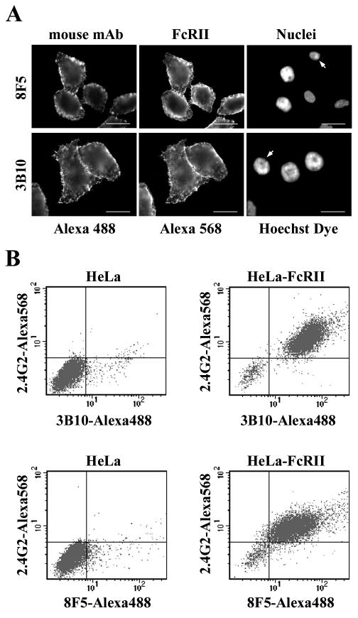
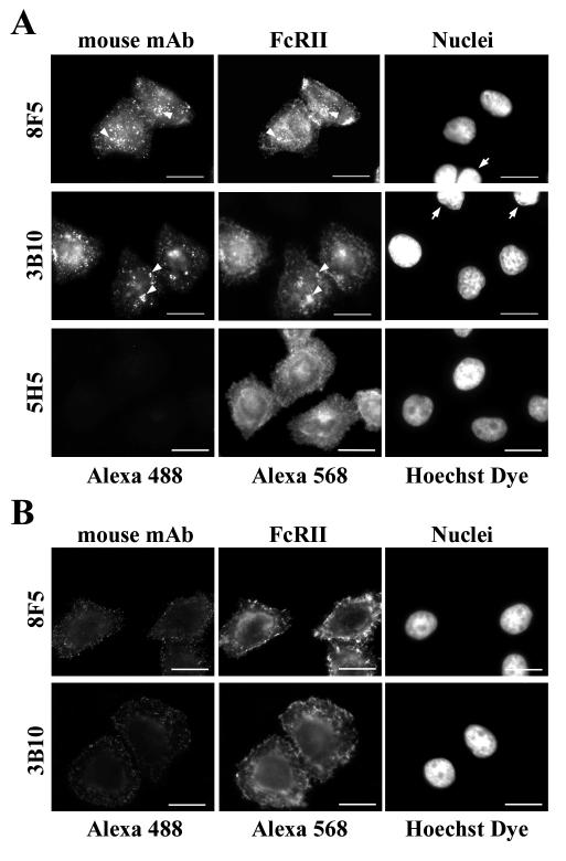
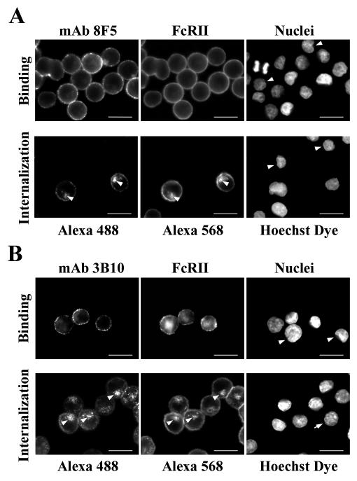
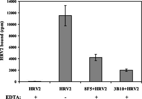
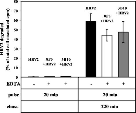
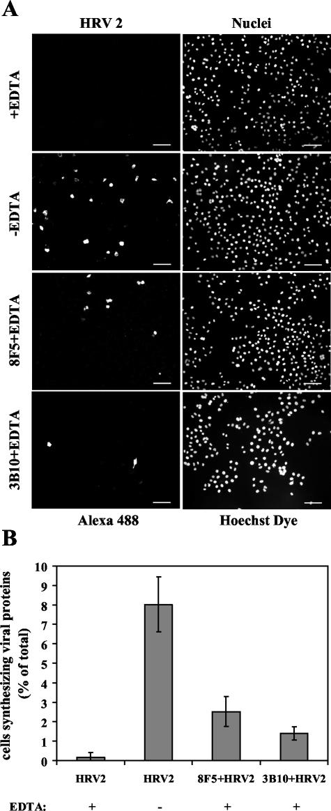
HeLa cells were stably transfected with a cDNA clone encoding the B1 isoform of the mouse FcgammaRII receptor (hereafter referred to as HeLa-FcRII cells). The receptor was expressed at high level at the plasma membrane in about 90% of the cells. These cells bound and internalized mouse monoclonal virus-neutralizing antibodies 8F5 and 3B10 of the subtype immunoglobulin G2a (IgG2a) and IgG1, respectively. Binding of the minor-group human rhinovirus type 2 (HRV2) to its natural receptors, members of the low-density lipoprotein receptor family, is dependent on the presence of Ca(2+) ions. Thus, chelating Ca(2+) ions with EDTA prevented HRV2 binding, entry, and infection. However, upon complex formation of (35)S-labeled HRV2 with 8F5 or 3B10, virus was bound, internalized, and degraded in HeLa-FcRII cells. Furthermore, challenge of these cells with HRV2-8F5 or HRV2-3B10 complexes resulted in de novo synthesis of viral proteins, as shown by indirect immunofluorescence microscopy. These data demonstrate that minor-group receptors can be replaced by surrogate receptors to mediate HRV2 cell entry, delivery into endosomal compartments, and productive uncoating. Consequently, the conformational change and uncoating of HRV2 appears to be solely triggered by the low-pH (pH </= 5.6) environment in these compartments.


Figures

FIG. 1.
FIG. 1.
FcRII expression in stably transfected HeLa-H1 cells. After transfection with pEF-Puro carrying the cDNA encoding murine FcRII and selection, clones were isolated and expanded. Cells grown on coverslips were incubated at 4°C with MAb 2.4G2 followed by Alexa 568-conjugated goat anti-rat IgG and viewed under a Zeiss Axiovert fluorescence microscope. Clone E1/1 is shown as an example. Comparison of the FcRII expression (A) and the nuclear staining of the same cells (B) shows that about 90% of the transfected cells express FcRII at the cell surface. Nuclei were stained with Hoechst dye. (C) At a higher magnification, plasma membrane staining can be seen. (D) FcRII expression in HeLa-FcRII cells grown in suspension. Bar, 100 μm in panels A and B; 20 μm in panels C and D.
FIG. 2.
FIG. 2.
MAbs 8F5 and 3B10 bind to FcRII. (A) HeLa-FcRII cells were grown on coverslips, incubated with 8F5 (40 μg/ml) or with 3B10 (15 μg/ml) at 4°C for 1 h, washed, fixed, and stained with Alexa 488-conjugated goat anti-mouse antibodies. Subsequently, cells were incubated with 2.4G2 followed by Alexa 568-conjugated goat anti-rat antibody. Nuclei were stained with Hoechst dye. Note the absence of Alexa 488 staining in cells that do not express FcRII (arrows). Bar, 20 μm. (B) Parental HeLa cells or HeLa-FcRII cells were incubated with 8F5 (40 μg/ml) or 3B10 (15 μg/ml) at 4°C for 1 h, washed, fixed, and stained with Alexa 488-conjugated goat anti-mouse antibodies followed by incubation with 2.4G2 and Alexa 568-conjugated goat anti-rat antibodies. Cells were analyzed by flow cytometry (for details, see Materials and Methods). One typical experiment out of three is shown. Note that only those cells that express FcRII bound the MAbs.
FIG. 3.
FIG. 3.
MAbs 8F5 and 3B10 are internalized into HeLa-FcRII cells. Cells were incubated at 34°C with 8F5 (40 μg/ml), 3B10 (15 μg/ml), or the control antibody 5H5 subtype IgG3 (40 μg/ml) for 60 min (8F5, 5H5) or for 20 min (3B10). Cells were washed, permeabilized (A) or nonpermeabilized (B), and processed for immunofluorescence microscopy as in Fig. 2. Note the perinuclear colocalization of MAbs and FcRII in permeabilized cells, indicative of localization in late endosomes (arrowheads), and the absence of any antibody uptake in FcRII-negative cells (right-hand panels, arrows). The control antibody 5H5 was not internalized by HeLa-FcRII cells. Bar, 20 μm.
FIG. 4.
FIG. 4.
Binding and internalization of antibodies is Ca2+ independent. HeLa-FcRII were grown in suspension and incubated with 8F5 (A) and 3B10 (B) in medium containing 10 mM EDTA either at 4°C for 1 h (binding, upper panels) or at 34°C for 60 min (8F5) or 20 min (3B10) (internalization, lower panels). Cells were washed, fixed, spun onto microscope slides and processed for indirect immunofluorescence microscopy as in Fig. 2. Upon internalization, colocalization of the antibodies with FcRII was observed (arrowheads). Again, cells devoid of FcRII do not bind and internalize antibodies (arrows). Bar, 20 μm.
FIG. 5.
FIG. 5.
HRV2 binds to HeLa-FcRII cells via MAbs in the absence of Ca2+. HeLa-FcRII suspension cells were incubated with 35S-labeled HRV2 alone or with HRV2 complexed to MAbs 8F5 and 3B10, respectively, in the presence or absence of 10 mM EDTA for 1 h at 4°C. Cells were pelleted and washed, and bound radioactivity was determined by liquid scintillation counting. Data given are means ± standard deviations (error bars) from one typical experiment out of three independent experiments, each carried out in triplicate.
FIG. 6.
FIG. 6.
Antibody-complexed virus is degraded in lysosomes. HeLa-FcRII cells grown in suspension were incubated for 20 min at 34°C with either 35S-HRV2 (in the absence of EDTA; control) or 35S-HRV2 complexed to 8F5 and 3B10, respectively, in the presence of EDTA to inhibit binding of the virus to its natural receptors. Cells were pelleted and washed, and incubation was continued for 220 min in normal medium. Total TCA-soluble and -precipitable counts were determined by liquid scintillation counting (see Materials and Methods). Degraded virus (TCA-soluble counts) is given as a percentage of cell-associated radioactivity after the 20-min incubation. The means ± standard deviations (error bars) of three independent experiments each carried out in triplicate are shown.
FIG. 7.
FIG. 7.
HeLa-FcRII cells are infected with antibody-complexed HRV2. (A) Cells were challenged with HRV2 (MOI, 10) in the presence or absence of 10 mM EDTA for 20 min at 34°C, washed, resuspended in infection medium, and further incubated for 18 h. Cells were fixed and stained for the presence of de novo-synthesized viral proteins by using MAb 8F5. Nuclei were stained with Hoechst dye. Images show 150 to 300 cells. Infection with HRV2-antibody complexes was carried out identically but in the presence of EDTA only. Bar, 100 μm. (B) Virus-synthesizing cells are given as percentage of the cells positive for viral antigen. One typical experiment out of three independent experiments is shown; results are presented as means ± standard deviations (error bars) of triplicates, with a total of about 1,500 to 2,000 cells examined.

References

    1. Arita, M., H. Horie, and A. Nomoto. 1999. Interaction of poliovirus with its receptor affords a high level of infectivity to the virion in poliovirus infections mediated by the Fc receptor. J. Virol. 73:1066-1074. - PMC - PubMed
    1. Bayer, N., D. Schober, M. Hüttinger, D. Blaas, and R. Fuchs. 2001. Inhibition of clathrin-dependent endocytosis has multiple effects on HRV2 cell entry. J. Biol. Chem. 276:3952-3962. - PubMed
    1. Bayer, N., D. Schober, E. Prchla, R. F. Murphy, D. Blaas, and R. Fuchs. 1998. Effect of bafilomycin A1 and nocodazole on endocytic transport in HeLa cells: implications for viral uncoating and infection. J. Virol. 72:9645-9655. - PMC - PubMed
    1. Brabec, M., G. Baravalle, D. Blaas, and R. Fuchs. 2003. Conformational changes, plasma membrane penetration, and infection by human rhinovirus type 2: role of receptors and low pH. J. Virol. 77:5370-5377. - PMC - PubMed
    1. Brown, M. S., J. Herz, and J. L. Goldstein. 1997. LDL receptor structure: calcium cages, acid baths and recycling receptors. Nature 388:629-630. - PubMed

Publication types

LinkOut - more resources