. 2004 Mar;78(6):2749-57.
doi: 10.1128/jvi.78.6.2749-2757.2004.

Cul7/p185/p193 binding to simian virus 40 large T antigen has a role in cellular transformation

Affiliations

Cul7/p185/p193 binding to simian virus 40 large T antigen has a role in cellular transformation

Syed Hamid Ali et al. J Virol. 2004 Mar.

Abstract

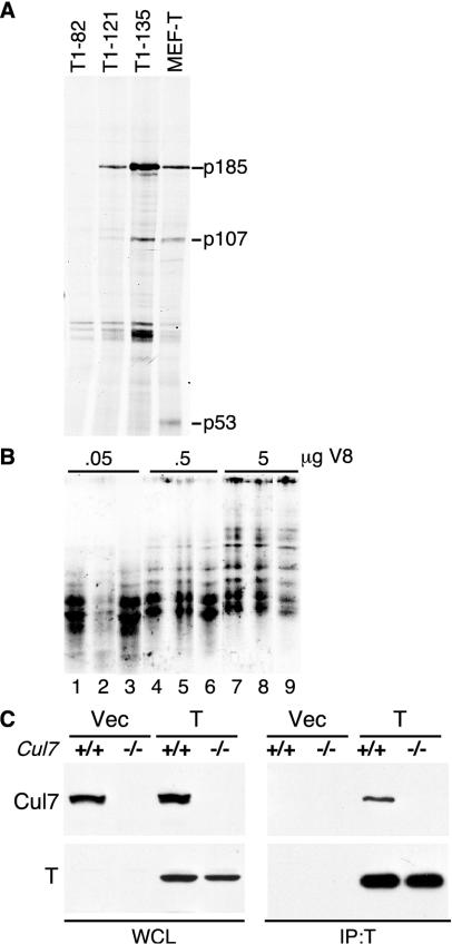
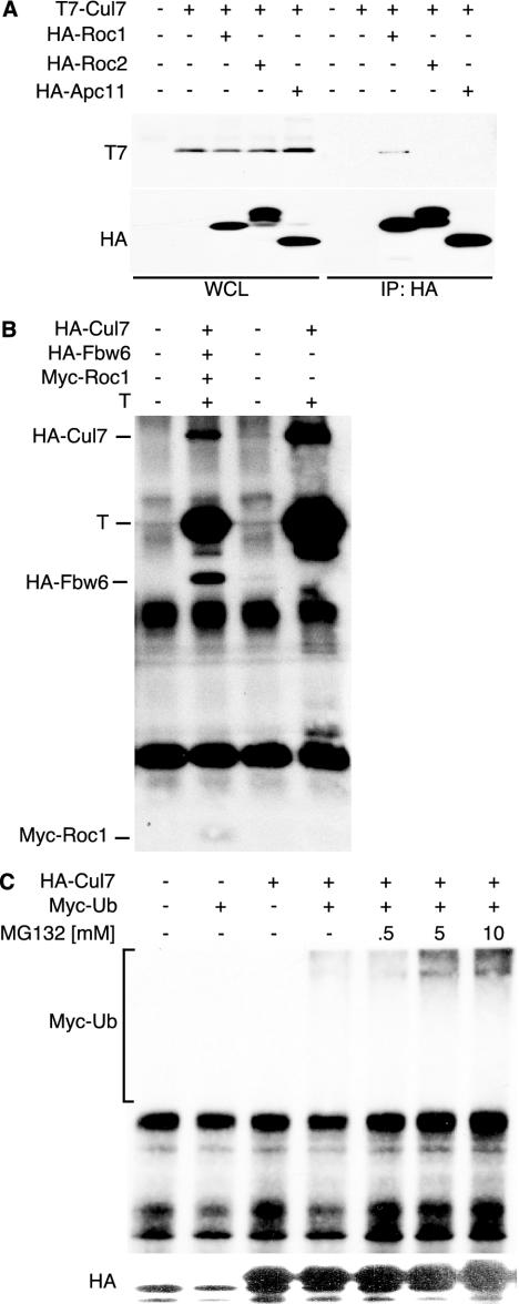
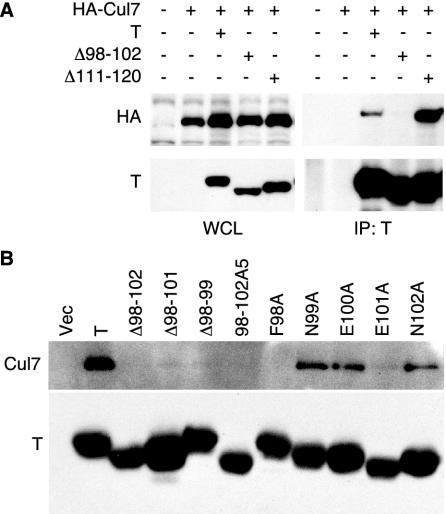
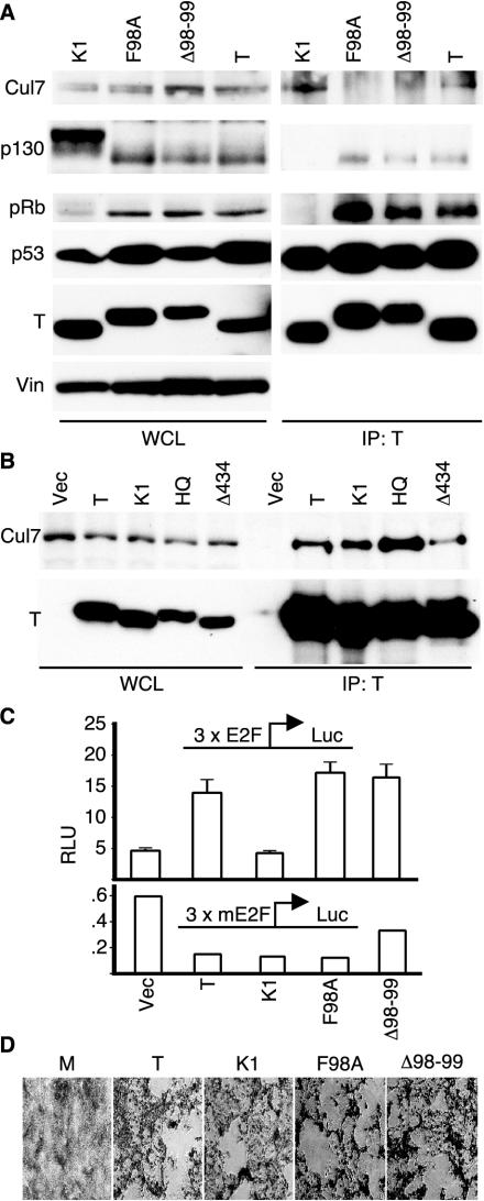
Simian virus 40 large T antigen (TAg) is a viral oncoprotein that can promote cellular transformation. TAg's transforming activity results in part by binding and inactivating key tumor suppressors, including p53 and the retinoblastoma protein (pRb). We have identified a TAg-associated 185-kDa protein that has significant homology to the cullin family of E3 ubiquitin ligases. TAg binds to an SCF-like complex that contains p185/Cul7, Rbx1, and the F box protein Fbw6. This SCF-like complex binds to an N-terminal region of TAg. Several p185/Cul7-binding-deficient mutants of TAg were generated that retained binding to pRb and p53 and were capable of overcoming Rb-mediated repression of E2F transcription. Despite binding to pRb and p53, these p185/Cul7-binding-defective mutants of TAg were unable to transform primary mouse embryo fibroblasts. Cells expressing p185/Cul7-binding-defective mutants of TAg were unable to grow to high density or grow in an anchorage-independent manner as determined by growth in soft agar. Considering the significance of other TAg-interacting proteins in regulation of the cell cycle, p185/Cul7 may also regulate an important growth control pathway.


Figures

FIG. 1.
FIG. 1.
Identification of p185/Cul7 in a TAg immuno-complex. (A) Lysates were prepared from 35S-labeled NIH 3T3 cells, incubated with purified N-terminal fragments of TAg or with unlabeled lysates from MEFs stably expressing wild-type TAg, and immunoprecipitated with TAg monoclonal antibody PAb419. A 185-kDa protein coprecipitated with full-length TAg and the fragments 1-121 and 1-135. TAg-associated proteins, p107 and p53, are identified. (B) The 185-kDa protein was in vitro translated from mouse p185 cDNA (lanes 1, 4, and 7) or immunoprecipitated from 35S-labeled NIH 3T3 cell lysates with either anti-p185 monoclonal antibody SA12 alone (lanes 2, 5, and 8) or with anti-TAg PAb419 in the presence of T1-135 (lanes 3, 6, and 9). The three samples were separated by SDS-PAGE, and the 185-kDa band was excised and digested with three different concentrations of V8 protease. The resulting peptides were separated in an SDS-PAGE and autoradiographed. (C) pBABE-puro-TAg or pBABE-vector (Vec) was stably expressed in wild-type (+/+) or Cul7 knockout (−/−) MEFs. Lysates were immunoprecipitated using anti-TAg antibody PAb419 and Western blotted with anti-p185/Cul7 monoclonal antibody SA12 (top panel) or anti-TAg monoclonal antibody PAb419 (bottom panel).
FIG. 2.
FIG. 2.
p185/Cul7 has properties of a cullin. (A) T7-tagged p185/Cul7 was transiently transfected with HA-tagged Roc1, Roc2, or APC11 into NIH 3T3 cells. Cell lysates were immunoprecipitated using anti-HA antibody 12CA5 and separated in an SDS-PAGE, followed by blotting with T7 and HA antibodies. (B) TAg was cotransfected with HA-p185/Cul7, HA-Fbw6, and myc-Roc1. The cell lysates were immunoprecipitated with anti-TAg (PAb419) antibody and blot-ted with anti-HA (12CA5), anti-TAg (PAb419), and anti-myc (9E10) antibodies. (C) HA-p185/Cul7 and myc-ubiquitin were transfected into NIH 3T3 cells. At 24 h after transfection, MG132 was added to the medium for 24 h. Lysates were immunoprecipitated with anti-HA antibody 12CA5, separated in an SDS-PAGE, and blotted with anti-HA (12CA5; top panel) or anti-myc (9E10; bottom panel) antibody.
FIG. 3.
FIG. 3.
TAg residues 98 to 102 are required for p185/Cul7 binding. (A) NIH 3T3 cells were transfected with HA-tagged Cul7 and wild-type TAg (T), deletion mutant Δ98-102, or Δ111-120. At 48 h following transfection, lysates were immunoprecipitated with TAg antibody PAb419, separated by SDS-PAGE, and blotted with PAb419 and HA antibody 12CA5. (B) Wild-type TAg and the indicated mutants of TAg within the region 98 to 102 were transfected in NIH 3T3 cells. Immunoprecipitation for TAg was followed by blotting with antibody against p185/Cul7 (SA12).
FIG. 4.
FIG. 4.
p185/Cul7 binding mutants of TAg. (A) Whole-cell lysates (left panels) of MEFs stably expressing wild-type TAg, F98A, Δ98-99, or K1 were separated in an SDS-PAGE or immunoprecipitated for TAg with PAb419 antibody (right panels). Western blotting was performed for p185/Cul7 (SA12), p130 (C20), pRb (XZ55), p53 (240), and TAg (PAb419), with vinculin (Vin) as a loading control. (B) NIH 3T3 cells were transiently transfected with TAg, K1, H42Q, or Δ434-444. Lysates were immunoprecipitated with PAb419 and blotted for p185/Cul7 (SA12; top panel) or TAg (PAb419; bottom). (C) NIH 3T3 cells were cotransfected with the indicated TAg plasmids and a reporter expressing the luciferase gene driven by a wild-type (3xE2F, upper panel) or mutated (3xmE2F, lower panel) promoter. To correctfor transfection efficiency, a third plasmid constitutively expressing the Renilla luciferase was included in all transfections. Cells were harvested 48 h following transfection, and the relative luciferase units were measured as described in Materials and Methods. The results are presented as the means of triplicates ± standard deviations. (D) CV-1P cells were transfected with wild-type SV40 genome or a mutant genome containing the K1, F98A, or Δ98-99 mutation. The cells were kept under observation for approximately 12 days until cytopathic effect was observed. When plaques became evident, the cultures were fixed and stained with crystal violet.
FIG. 5.
FIG. 5.
TAg transformation of MEFs. (A) Equal numbers of primary MEFs stably expressing wild-type TAg (open circle), K1 (square), F98A (closed triangle), or Δ98-99 (closed circle) were seeded in 60-mm dishes. The cells were fed on alternate days with medium containing either 10% or 1% FCIS. The results in the top two and lower two panels are from two independently generated pools of stably transfected MEFs. The results are presented as means of total number of cells (105) on four 60-mm plates ± standard deviations. (B) An equal number of the above-mentioned MEFs was seeded in soft agar and allowed to grow for 3 weeks. Clusters of cells containing four or more cells were scored as colonies. Total colonies and single cells were counted. The results are shown as the percentage of colonies observed for each TAg clone. Results from two separate pools (i and ii) of F98A and Δ98-99 MEFs are presented.

References

    1. Ali, S. H., and J. A. DeCaprio. 2001. Cellular transformation by SV40 large T antigen: interaction with host proteins. Semin. Cancer Biol. 11:15-23. - PubMed
    1. Arai, T., J. S. Kasper, J. R. Skaar, S. H. Ali, C. Takahashi, and J. A. DeCaprio. 2003. Targeted disruption of p185/Cul7 gene results in abnormal vascular morphogenesis. Proc. Natl. Acad. Sci. USA 100:9855-9860. - PMC - PubMed
    1. Avantaggiati, M. L., M. Carbone, A. Graessmann, Y. Nakatani, B. Howard, and A. S. Levine. 1996. The SV40 large T antigen and adenovirus E1a oncoproteins interact with distinct isoforms of the transcriptional co-activator, p300. EMBO J. 15:2236-2248. - PMC - PubMed
    1. Bai, C., P. Sen, K. Hofmann, L. Ma, M. Goebl, J. W. Harper, and S. J. Elledge. 1996. SKP1 connects cell cycle regulators to the ubiquitin proteolysis machinery through a novel motif, the F-box. Cell 86:263-274. - PubMed
    1. Bargonetti, J., P. N. Friedman, S. E. Kern, B. Vogelstein, and C. Prives. 1991. Wild-type but not mutant p53 immunopurified proteins bind to sequences adjacent to the SV40 origin of replication. Cell 65:1083-1091. - PubMed

Publication types