. 2004 Mar;78(6):2790-807.
doi: 10.1128/jvi.78.6.2790-2807.2004.

Genetic and phenotypic analyses of human immunodeficiency virus type 1 escape from a small-molecule CCR5 inhibitor

Affiliations

Genetic and phenotypic analyses of human immunodeficiency virus type 1 escape from a small-molecule CCR5 inhibitor

Shawn E Kuhmann et al. J Virol. 2004 Mar.

Erratum in

  • J Virol. 2005 Jun;78(12):6706

Abstract

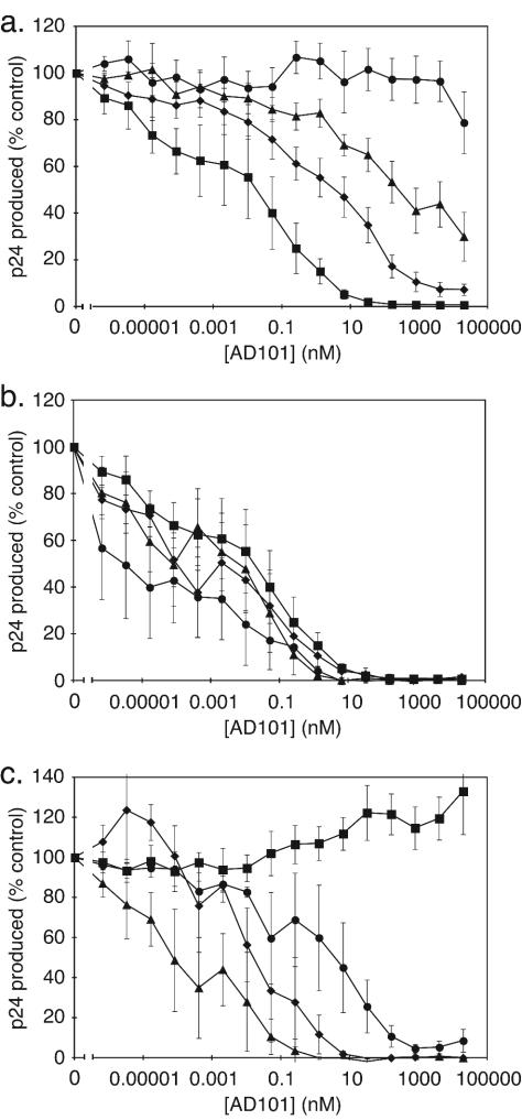
We have described previously the generation of an escape variant of human immunodeficiency virus type 1 (HIV-1), under the selection pressure of AD101, a small molecule inhibitor that binds the CCR5 coreceptor (A. Trkola, S. E. Kuhmann, J. M. Strizki, E. Maxwell, T. Ketas, T. Morgan, P. Pugach, S. X. L. Wojcik, J. Tagat, A. Palani, S. Shapiro, J. W. Clader, S. McCombie, G. R. Reyes, B. M. Baroudy, and J. P. Moore, Proc. Natl. Acad. Sci. USA 99:395-400, 2002). The escape mutant, CC101.19, continued to use CCR5 for entry, but it was at least 20,000-fold more resistant to AD101 than the parental virus, CC1/85. We have now cloned the env genes from the the parental and escape mutant isolates and made chimeric infectious molecular clones that fully recapitulate the phenotypes of the corresponding isolates. Sequence analysis of the evolution of the escape mutants suggested that the most relevant changes were likely to be in the V3 loop of the gp120 glycoprotein. We therefore made a series of mutant viruses and found that full AD101 resistance was conferred by four amino acid changes in V3. Each change individually caused partial resistance when they were introduced into the V3 loop of a CC1/85 clone, but their impact was dependent on the gp120 context in which they were made. We assume that these amino acid changes alter how the HIV-1 Env complex interacts with CCR5. Perhaps unexpectedly, given the complete dependence of the escape mutant on CCR5 for entry, monomeric gp120 proteins expressed from clones of the fully resistant isolate failed to bind to CCR5 on the surface of L1.2-CCR5 cells under conditions where gp120 proteins from the parental virus and a partially AD101-resistant virus bound strongly. Hence, the full impact of the V3 substitutions may only be apparent at the level of the native Env complex.


Figures

FIG. 1.
FIG. 1.
Infectious molecular clones recapitulate the phenotype of the AD101-resistant escape mutant CC101.19. (a) Chimeric molecular clones containing env genes derived from the parental CC1/85 isolate (open symbols) or the passage 19 escape mutant (solid symbols) were cultured in primary CD4+ T cells in the presence of the AD101 concentrations indicated, and the extent of HIV-1 replication was determined. The individual clones are designated CC1/85 cl.6 (open squares), CC1/85 cl.7 (open circles), CC1/85 cl.8 (open triangles), CC101.19 cl.3 (solid squares), CC101.19 cl.7 (solid circles), and CC101.19 cl.15 (solid triangles). (b to d) Four of the above clones (same symbols) were also tested for sensitivity to SCH-C (b), RANTES (c), and PA14 (d). Panel a displays data from a single representative experiment. Panels b to d show data averaged from three independent experiments, with the error bars indicating the standard error of the mean (SEM).
FIG. 2.
FIG. 2.
Phylogenetic analysis of gp120 sequences from CC1/85 and related viruses. Sequence diversity is represented by horizontal distance. CC1/85 sequences are represented by black bars. Sequences from the CCcon.4 (passage 4), CCcon.10 (passage 10), and CCcon.20 (passage 20) isolates are shown by blue bars, with the darker bars denoting the later isolates. The sequences from viruses isolated under AD101 selection pressure are represented in yellow, orange, and red. The sequences in green represent in vivo diversification that generates the CC2/86, CC7/86, and CC12/86 isolates. The CC101.22R2, CC101.22R5, and CC101.22R9 sequences in different shades of purple are derived from escape mutant viruses cultured in the absence of AD101 for a further nine passages.
FIG.3.
FIG.3.
V3 sequence alignments of Env clones. The first line shows the consensus V3 sequence of the CC1/85 isolate based on eight clones. The locations of specific amino acids and their corresponding amino acid numbers, based on the HXBc2 sequence, are shown above the consensus line. There are five blocks of sequence data below the consensus line. Each block includes the following information (from left to right): N, the number of clones from a given isolate having the indicated V3 sequence; the source isolate, i.e., the isolate, based on the nomenclature described in the text, from which the clones were generated; and the sequence. The sequence is shown in reference to the consensus, with a dash representing identity to the consensus. Where one or more clones differ from the consensus, the amino acid found in that clone or clones is shown. Block 1 contains the eight clones from the CC1/85 isolate, seven of which are identical to the consensus and one of which contains the H308P polymorphism. This polymorphism and others discussed in the main text are highlighted in red. Block 2 contains select isolates generated from the cultures treated with AD101; the clones are shown first in order of the isolate from which they were derived and then in order of the frequency at which each clone is present in that isolate. Block 3 contains clones derived from isolates cultured for 22 passages before AD101 was removed for an additional 9 passages. Block 4 contains clones from three control isolates passaged in the absence of AD101. Block 5 contains clones from isolates derived from patient Case C at 13 (CC2/86), 18 (CC7/86), and 23 (CC12/86) months after the CC1/85 isolate. The $ symbol in one CC101.14 clone indicates the position of a premature stop codon.
FIG. 4.
FIG. 4.
Alignments of the consensus gp160 sequences from critical time points in the AD101 escape process. The consensus amino acid sequence from the parental isolate, CC1/85, is given on the top line. Capital letters indicate that there was identity among all eight of the aligned clones. Lowercase letters indicate the amino acid sequence of most but not all of the clones. The question mark indicates either that there was a tie between amino acids at a given position or that the majority of clones had a gap at this position. A gap found in all clones is indicated by a dot. Aligned with the CC1/85 consensus are the consensus sequences from isolates from passages 4, 10, and 20 (CC101.4, CC101.10, and CC101.20, respectively), derived under AD101 selection pressure. Also depicted are consensus sequences from the CC101.22R9 isolate, which was cultured in the absence of AD101 for 9 passages after 22 passages in the presence of AD101; the passage control CCcon.20 isolate, which was cultured for 20 passages in the same PBMC as the AD101-treated cultures, but in the absence of drug; and CC12/86, which was isolated from individual Case C 23 months after the CC1/85 isolate was obtained. If the aligned sequence is identical to that of the CC1/85 consensus line, this is indicated by a dash; otherwise, the symbols described above are used to depict differences from the consensus sequence. Amino acids highlighted in red are those that were selected for in the AD101-treated cultures but not in the control cultures or in vivo. Amino acids highlighted in green are those that were selected for in both the AD101-treated and control cultures. Amino acids highlighted in blue are those that were selected for in the AD101-treated cultures and in vivo but not in the control cultures. The numbering system is based on that of HXBc2. The amino acid under the "0” of each number is the one intended to be indicated by the label. The variable and constant regions of gp120, and the gp120-gp41 cleavage site, are also indicated. The outlined amino acids are those which have been implicated in the binding of, or neutralization by, MAb 17b (59, 94, 105, 118). Amino acids on a gray background are those implicated in forming part of a conserved coreceptor binding site that partially overlaps the epitope for 17b (93, 94).
FIG. 5.
FIG. 5.
AD101 sensitivity of NL4-3/env chimeric viruses with interchanged C2, V3, and C3 regions. The AD101-sensitive clones CC1/85 cl.7 (squares) and AD101-resistant CC101.19 cl.7 (diamonds) and the chimeric clones CC1/85 cl.7(8) (triangles) and CC101.19 cl.7(8) (circles) were all tested for AD101 sensitivity in an assay of HIV-1 replication in primary CD4+ T cells. The results depicted are the average of four or five independent experiments, with the error bars indicating the SEM.
FIG. 6.
FIG. 6.
Effect of specific amino acid substitutions in the V3 loop on sensitivity to AD101. (a) The parental CC1/85 cl.7 virus (squares) and mutants CC1/85 cl.7(H308P) (diamonds), CC1/85 cl.7(K305R, H308P) (triangles), and CC1/85 cl.7(K305R, H308P, A316V, G321E) (circles) were all tested for AD101 sensitivity in an assay of HIV-1 replication in primary CD4+ T cells. (b) Three additional mutants derived from CC1/85 cl.7 (squares) were also tested for AD101 sensitivity: CC1/85 cl.7(K305R) (diamonds), CC1/85 cl.7(A316V, G321E) (triangles), and CC1/85 cl.7(K305R, A316V, G321E) (circles). (c) Reverse mutants made in the context of the AD101-resistant parental clone CC101.19 cl.7 (squares) were evaluated: CC101.19 cl.7(R305K, P308H, V316A, E321G) (diamonds), CC101.19 cl.7(P308H) (triangles), and CC101.19 cl.7(R305K, P308H) (circles). The results depicted in all panels are the average of four or five independent experiments, with the error bars indicating the SEM.
FIG. 7.
FIG. 7.
Binding of monomeric gp120s from AD101-sensitive and -resistant viruses to L1.2-CCR5 cells in the presence and absence of AD101. The monomeric gp120 proteins indicated (100 μg/ml) with or without biotinylated CD4-lgG2 (100 μg/ml) were added to L1.2-CCR5 cells with and without AD101 (1 μM). The extent of binding of the gp120-CD4-IgG2 complex was measured as the mean fluorescence intensity (MFI) and is shown in arbitrary units (A.U.). Uncorrected mean fluorescence intensity values from a single, representative experiment are presented. The intrinsic fluorescence in the absence of gp120 is indicated in the “no gp120” column.

References

    1. Aarons, E. J., S. Beddows, T. Willingham, L. Wu, and R. A. Koup. 2001. Adaptation to blockade of human immunodeficiency virus type 1 entry imposed by the anti-CCR5 monoclonal antibody 2D7. Virology 287:382-390. - PubMed
    1. Adachi, A., H. E. Gendelman, S. Koenig, T. Folks, R. Willey, A. Rabson, and M. A. Martin. 1986. Production of acquired immunodeficiency syndrome-associated retrovirus in human and nonhuman cells transfected with an infectious molecular clone. J. Virol. 59:284-291. - PMC - PubMed
    1. Allaway, G. P., K. L. Davis-Bruno, G. A. Beaudry, E. B. Garcia, E. L. Wong, A. M. Ryder, K. W. Hasel, M. C. Gauduin, R. A. Koup, J. S. McDougal, and P. J. Maddon. 1995. Expression and characterization of CD4-IgG2, a novel heterotetramer that neutralizes primary HIV type 1 isolates. AIDS Res. Hum. Retrovir. 11:533-539. - PubMed
    1. Altschul, S. F., W. Gish, W. Miller, E. W. Myers, and D. J. Lipman. 1990. Basic local alignment search tool. J. Mol. Biol. 215:403-410. - PubMed
    1. Alves, K., M. Canzian, and E. L. Delwart. 2002. HIV type 1 envelope quasispecies in the thymus and lymph Nodes of AIDS patients. AIDS Res. Hum. Retrovir. 18:161-165. - PubMed

Publication types

MeSH terms

Associated data

LinkOut - more resources