. 2004 Mar;78(6):2948-55.
doi: 10.1128/jvi.78.6.2948-2955.2004.

RNA recombination plays a major role in genomic change during circulation of coxsackie B viruses

Affiliations

RNA recombination plays a major role in genomic change during circulation of coxsackie B viruses

M Steven Oberste et al. J Virol. 2004 Mar.

Abstract

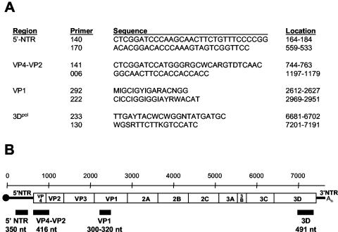
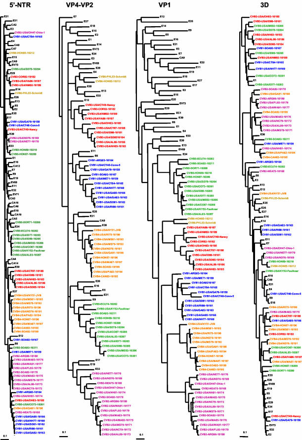
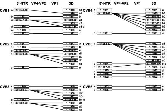
RNA recombination has been shown to occur during circulation of enteroviruses, but most studies have focused on poliovirus. To examine the role of recombination in the evolution of the coxsackie B viruses (CVB), we determined the partial sequences of four genomic intervals for multiple clinical isolates of each of the six CVB serotypes isolated from 1970 to 1996. The regions sequenced were the 5'-nontranslated region (5'-NTR) (350 nucleotides [nt]), capsid (VP4-VP2, 416 nt, and VP1, approximately 320 nt), and polymerase (3D, 491 nt). Phylogenetic trees were constructed for each genome region, using the clinical isolate sequences and those of the prototype strains of all 65 enterovirus serotypes. The partial VP1 sequences of each CVB serotype were monophyletic with respect to serotype, as were the VP4-VP2 sequences, in agreement with previously published studies. In some cases, however, incongruent tree topologies suggested that intraserotypic recombination had occurred between the sequenced portions of VP2 and VP1. Outside the capsid region, however, isolates of the same serotype were not monophyletic, indicating that recombination had occurred between the 5'-NTR and capsid, the capsid and 3D, or both. Almost all clinical isolates were recombinant relative to the prototype strain of the same serotype. All of the recombination partners appear to be members of human enterovirus species B. These results suggest that recombination is a frequent event during enterovirus evolution but that there are genetic restrictions that may influence recombinational compatibility.


Figures

FIG. 1.
FIG. 1.
Schematic representation of the enterovirus genome and primers used for RT-PCR amplification. (A) Oligonucleotide primer pairs used for amplification of four genomic intervals of each of the coxsackie B virus isolates. Nucleotide sequence coordinates are relative to the genome of poliovirus 1, Mahoney strain. Primer 006 is a modification of primer 5− (11, 23); the use of primers 292 and 222 for molecular serotyping was described previously (21, 24); primers 233 and 130 are the same as primers 3D+ and 3D−, respectively (27). (B) The approximate location and size of PCR products are shown under the enterovirus genetic map. The scale, in nucleotides, is indicated above the map.
FIG.2.
FIG.2.
Phylogenetic relationships of CVB clinical isolates and other members of HEV-B. Trees were constructed by the neighbor-joining method using DNADist and Neighbor programs (PHYLIP) (7). Trees for the different genomic intervals were plotted to the same scale, indicated by the scale bar below and to the left of each tree. The CVB isolates are color coded by serotype; isolates of other serotypes are shown in black. Because the major clusters radiate from a multifurcating node at the center of the tree, their order in the tree is arbitrary.
FIG. 3.
FIG. 3.
Summary of the different recombination patterns identified for each CVB serotype. Bars represent phylogenetically distinct sequences (i.e., a monophyletic lineage of homologous serotype), defined by intervening isolates of a heterologous serotype in the trees in Fig. 2. Lowercase letters indicate the 5′-NTR lineage within each serotype. Numbers after the lowercase letters indicate the 3D sublineage of that 5′-NTR lineage. The lineages are labeled chronologically, except when their relationship in 3D dictates a different order to maintain clarity. Numbers in the bars are the number of isolates of that lineage and the range of years in which the lineage was isolated.

References

    1. Andersson, P., K. Edman, and A. M. Lindberg. 2002. Molecular analysis of the echovirus 18 prototype. Evidence of interserotypic recombination with echovirus 9. Virus Res. 85:71-83. - PubMed
    1. Brown, B. A., K. Maher, M. S. Oberste, and M. A. Pallansch. 2003. Complete genomic sequencing shows that polioviruses and members of human enterovirus species C are closely related in the non-capsid coding region. J. Virol. 77:8973-8984. - PMC - PubMed
    1. Cammack, N., A. Phillips, G. Dunn, V. Patel, and P. D. Minor. 1988. Intertypic genomic rearrangements of poliovirus strains in vaccinees. Virology 167:507-514. - PubMed
    1. Cherkasova, E. A., E. A. Korotkova, M. L. Yakovenko, O. E. Ivanova, T. P. Eremeeva, K. M. Chumakov, and V. I. Agol. 2002. Long-term circulation of vaccine-derived poliovirus that causes paralytic disease. J. Virol. 76:6791-6799. - PMC - PubMed
    1. Cuervo, N. S., S. Guillot, N. Romanenkova, M. Combiescu, A. Aubert-Combiescu, M. Seghier, V. Caro, R. Crainic, and F. Delpeyroux. 2001. Genomic features of intertypic recombinant Sabin poliovirus strains excreted by primary vaccinees. J. Virol. 75:5740-5751. - PMC - PubMed