. 2004 Mar;78(6):2984-93.
doi: 10.1128/jvi.78.6.2984-2993.2004.

US11 of herpes simplex virus type 1 interacts with HIPK2 and antagonizes HIPK2-induced cell growth arrest

Affiliations

US11 of herpes simplex virus type 1 interacts with HIPK2 and antagonizes HIPK2-induced cell growth arrest

Stéphane Giraud et al. J Virol. 2004 Mar.

Abstract

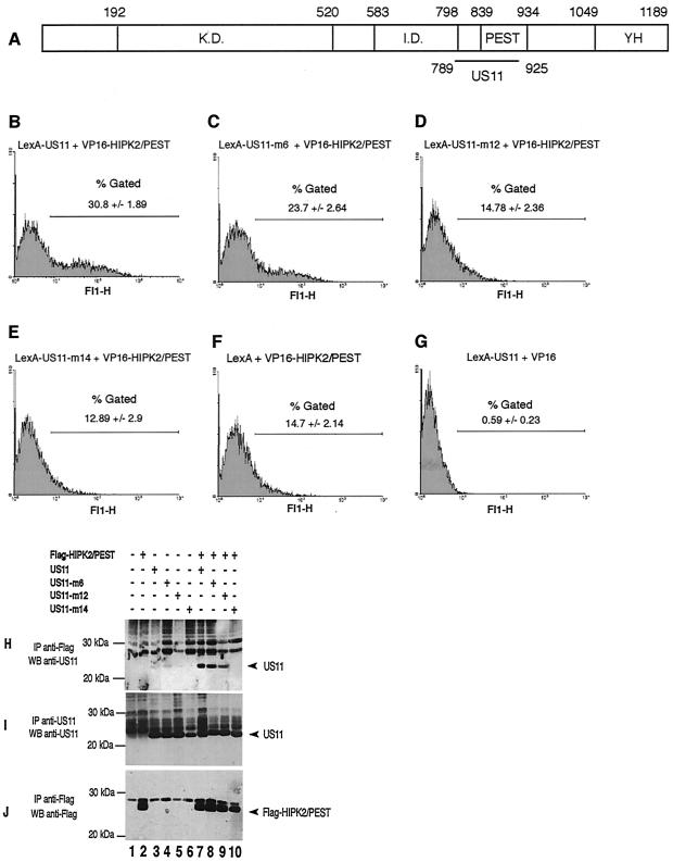
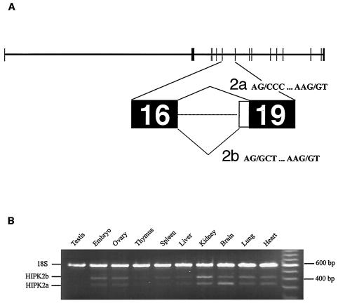
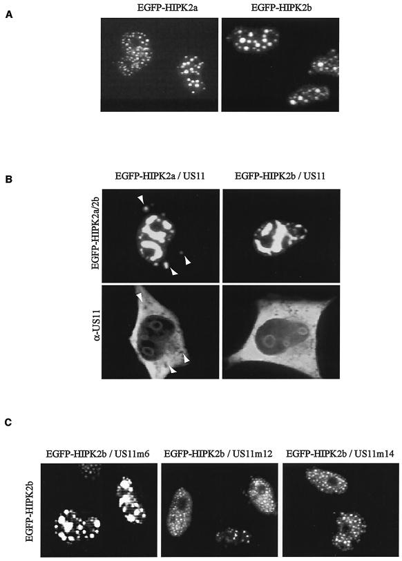
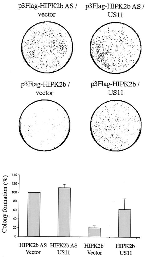
Homeodomain-interacting protein kinase 2 (HIPK2) is a nuclear serine/threonine kinase of the subfamily of dual-specificity Yak1-related kinase proteins. HIPK2 was first described as a homeodomain-interacting protein kinase acting as a corepressor for homeodomain transcription factors. More recently, it was reported that HIPK2 plays a role in p53-mediated cellular apoptosis and could also participate in the regulation of the cell cycle. US11 protein of herpes simplex virus type 1 is a multifunctional protein involved in the regulation of several processes related to the survival of cells submitted to environmental stresses by mechanisms that are not fully elucidated. In an attempt to better understand the multiple functions of US11, we identified cellular binding partners of this protein by using the yeast two-hybrid system. We report that US11 interacts with HIPK2 through the PEST domain of HIPK2 and that this interaction occurs also in human cells. This interaction modifies the subcellular distribution of HIPK2 and protects the cell against the HIPK2-induced cell growth arrest.


Figures

FIG. 1.
FIG. 1.
(A) Schematic representation of the HIPK2 coding sequence. K.D., kinase domain; I.D., interaction domain for homeoproteins; PEST, proline-, glutamic acid-, serine-, and threonine-rich domain; YH, tyrosine- and histidine-rich domain. Numbers indicate amino acid positions. The horizontal black line marks the delineation of the interaction domain of HIPK2 with US11. (B to G) Interaction of LexA-US11 and LexA-US11 mutant forms with VP16-HIPK2/PEST. The protein-protein interaction was measured by the yeast two-hybrid analyses with the GFP gene as a reporter. Yeast strain YRN974 with an integrated LexA operator-GFP gene was used for these analyses. (B) Yeast cells were transformed with expression vectors encoding LexA-US11 and VP16-HIPK2/PEST proteins. (C to E) Yeast cells were transformed with expression vectors encoding VP16-HIPK2/PEST combined with various vectors coding for different LexA-US11 mutants, i.e., LexA-US11-m6, LexA-US11-m12, and LexA-US11-m14. Mutations carried by these proteins are detailed in reference . (F) Yeast cells expressing LexA alone and VP-16-HIPK2/PEST. (G) Yeast cells expressing LexA-US11 and VP-16 alone. (H to J) Interaction of the PEST domain of HIPK2 with US11 and US11 mutants in human cells. HeLa cells were cotransfected with empty vectors (lane 1) or the indicated expression vectors containing a cDNA encoding Flag-HIPK2/PEST (lanes 2 and 7 to 10), US11 (lanes 3 and 7), US11-m6 (lanes 4 and 8), US11-m12 (lanes 5 and 9), or US11-m14 (lanes 6 and 10). Cell lysates were subjected to immunoprecipitation (IP) with an anti-Flag antibody (H and J) or an anti-US11 antibody (I). Purified proteins were analyzed by Western blotting with an anti-US11 (H and I) or an anti-Flag (J) antibody. Arrowheads indicate the positions of US11 (H and I) or Flag-HIPK2/PEST (J). Molecular masses are shown on the left in kilodaltons. +, present; −, absent.
FIG. 2.
FIG. 2.
(A) Structural organization of the murine HIPK2 gene. The bioinformatic analysis is described in Materials and Methods. Exons are represented by filled boxes. Potential alternative splicing occurring between exons 16 and 19 according to the numbering of model maker software (http://www.ncbi.nlm.nih.gov/mapview) are labeled 2a and 2b. (B) Analysis of the HIPK2 gene expression in different murine tissues by RT-PCR. Total mRNA (Clontech) was reverse transcribed, and the HIPK2a and HIPK2b cDNA and 18S rRNA were amplified with primers described in Materials and Methods. Positions of the DNA fragments corresponding to 18S, HIPK2b, and HIPK2a are indicated to the left of the figure. Sizes of the DNA fragments are indicated to the right of the figure in base pairs.
FIG. 3.
FIG. 3.
(A) Intracellular localization of HIPK2a and HIPK2b. EGFP-HIPK2a and EGFP-HIPK2b expression vectors were transiently expressed in HeLa cells. The EGFP fluorescence signal was visualized 48 h after transfection. (B) Intracellular distribution of HIPK2a and HIPK2b when coexpressed with US11. US11 disrupts the nuclear localization of HIPK2a and HIPK2b. HeLa cells were transfected with expression vectors encoding US11 protein and a vector encoding either EGFP-HIPK2a or EGFP-HIPK2b. At 48 h after transfection, cells were fixed and stained with an anti-US11 antibody and Texas Red-conjugated anti-rabbit IgG. Arrows indicate HIPK2a found in the cytoplasm. (C) Intracellular distribution of HIPK2b when coexpressed with different US11 mutants. HeLa cells were transfected with expression vector encoding EGFP-HIPK2b combined with expression vector coding for either US11-m6, US11-m12, or US11-m14. The EGP-HIPK2b fluorescence signal was visualized 48 h after transfection.
FIG. 4.
FIG. 4.
Kinase activity of HIPK2b. HEK 293 cells were transfected with either 3Flag-HIPK2b, Flag-K221R/HIPK2, or 3Flag expression vector. The corresponding proteins were immunoprecipitated with anti-Flag antibody, and their activities were determined by immune kinase complex assay with GST, GST-US11-m55, or GST-US11 as a substrate. The figure shows an autoradiogram of proteins separated by SDS-PAGE. Lanes 1 to 5, experiment performed with 3Flag-HIPK2b expression vector; lane 6, experiment performed with Flag-K221R/HIPK2 expression vector; lanes 7 to 11, experiment performed with 3Flag expression vector. Lanes 4 and 5 contain, respectively, 1 and 2 μg of GST-US11. Positions of the radiolabeled proteins are indicated to the left of the figure. Molecular masses are indicated to the right of the figure. +, present; −, absent.
FIG. 5.
FIG. 5.
HIPK2 cell growth arrest antagonized by US11 expression. HEK 293 cells were transfected with plasmids p3Flag-HIPK2b and p3Flag-HIPK2b AS combined with either expression vector for US11 or the corresponding empty vector. At 24 h after transfection, cells were selected with G418 and surviving colonies were stained with crystal violet. The combination of the vectors used for transfection is indicated above each corresponding box. Colony formation with the control vector p3Flag-HIPK2b AS was set as 100%. Results are means ± standard deviations of the results from three independent experiments (lower panel).

Similar articles

Cited by

References

    1. Besse, S., J.-J. Diaz, E. Pichard, K. Kindbeiter, J.-J. Madjar, and F. Puvion-Dutilleul. 1996. In situ hybridization and immuno-electron microscope analyses of the Us11 gene of herpes simplex virus type 1 during transient expression. Chromosoma 104:434-444. - PubMed
    1. Bhat, R. A., and B. Thimmappaya. 1983. Two small RNAs encoded by Epstein-Barr virus can functionally substitute for the virus-associated RNAs in the lytic growth of adenovirus 5. Proc. Natl. Acad. Sci. USA 80:4789-4793. - PMC - PubMed
    1. Brown, S. M., and J. Harland. 1987. Three mutants of herpes simplex virus type 2: one lacking the genes Us10, Us11, Us12 and two in which Rs has been extended by 6 kb to 0, 91 map units with loss of Us sequences between 0, 94 and the Us/TRs junction. J. Gen. Virol. 68:1-18. - PubMed
    1. Cassady, K. A., and M. Gross. 2002. The herpes simplex virus type 1 U(S)11 protein interacts with protein kinase R in infected cells and requires a 30-amino-acid sequence adjacent to a kinase substrate domain. J. Virol. 76:2029-2035. - PMC - PubMed
    1. Catez, F., M. Erard, N. Schaerer-Uthurralt, K. Kindbeiter, J.-J. Madjar, and J.-J. Diaz. 2002. A unique motif for nucleolar retention and nuclear export regulated by phosphorylation. Mol. Cell. Biol. 22:1126-1139. - PMC - PubMed

Publication types

MeSH terms

Associated data