Comparative Study
. 2004 Mar;78(6):3162-9.
doi: 10.1128/jvi.78.6.3162-3169.2004.

Inhibition of different Lassa virus strains by alpha and gamma interferons and comparison with a less pathogenic arenavirus

Affiliations
Comparative Study

Inhibition of different Lassa virus strains by alpha and gamma interferons and comparison with a less pathogenic arenavirus

Marcel Asper et al. J Virol. 2004 Mar.

Abstract

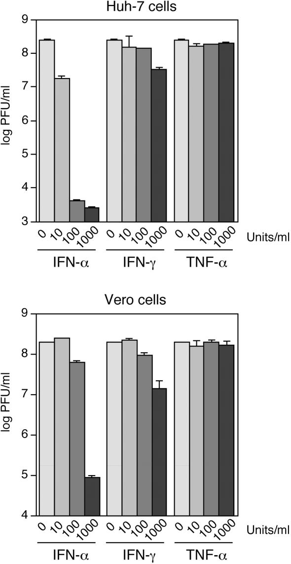
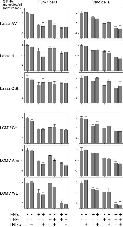
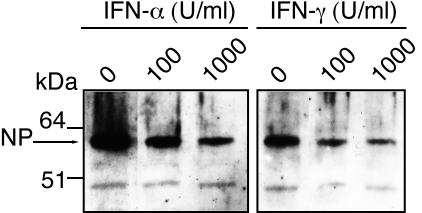
The high pathogenicity of Lassa virus is assumed to involve resistance to the effects of interferon (IFN). We have analyzed the effects of alpha IFN (IFN-alpha), IFN-gamma, and tumor necrosis factor alpha (TNF-alpha) on replication of Lassa virus compared to the related, but less pathogenic, lymphocytic choriomeningitis virus (LCMV). Three low-passage Lassa virus strains (AV, NL, and CSF), isolated from humans with mild to fulminant Lassa fever, were tested. Lassa virus replication was inhibited by IFN-alpha and IFN-gamma, but not TNF-alpha, in Huh7 and Vero cells. The degree of IFN sensitivity of a Lassa virus isolate did not correlate with disease severity in human patients. Furthermore, cytokine effects observed for Lassa virus and LCMV (strains CH-5692, Armstrong, and WE) were similar. To address the mechanisms involved in the IFN effect, we used cell lines in which overexpression of IFN-stimulated proteins promyelocytic leukemia protein (PML) and Sp100 could be induced. Both proteins reside in PML bodies, a cellular target of the LCMV and Lassa virus Z proteins. Overexpression of PML or Sp100 did not affect replication of either virus. This, together with the previous finding that PML knockout facilitates LCMV replication in vitro and in vivo (M. Djavani, J. Rodas, I. S. Lukashevich, D. Horejsh, P. P. Pandolfi, K. L. Borden, and M. S. Salvato, J. Virol. 75:6204-6208, 2001; W. V. Bonilla, D. D. Pinschewer, P. Klenerman, V. Rousson, M. Gaboli, P. P. Pandolfi, R. M. Zinkernagel, M. S. Salvato, and H. Hengartner, J. Virol. 76:3810-3818, 2002), describes PML as a mediator within the antiviral pathway rather than as a direct effector protein. In conclusion, the high pathogenicity of Lassa virus compared to LCMV is probably not due to increased resistance to the effects of IFN-alpha or IFN-gamma. Both cytokines inhibit replication which is relevant for the design of antiviral strategies against Lassa fever with the aim of enhancing the IFN response.


Figures

FIG. 1.
FIG. 1.
Sensitivity of VSV to different concentrations of human IFN-α, IFN-γ, and TNF-α in Huh7 and Vero cells. Cells were infected at an MOI of 0.01, and the virus titer was determined in the supernatant 24 h later by plaque assay. The values for two experiments are shown on a logarithmic scale.
FIG. 2.
FIG. 2.
Growth kinetics of Lassa virus AV. Vero cells were infected at different MOIs, and the concentration of S RNA molecules (top graph) and infectious particles (bottom graph) in supernatant was measured by real-time PCR and an immunological focus assay, respectively. The table at the top of the figure shows the relation between both measurements, expressed as S RNA molecules per PFU.
FIG. 3.
FIG. 3.
Sensitivity of Lassa virus and LCMV to IFN-α, IFN-γ, and TNF-α. Huh7 and Vero cells were preincubated with human IFN-α, IFN-γ, or TNF-α (1,000 U/ml) (+) or combinations thereof for 24 h before virus infection or left untreated. Cells were infected with Lassa virus strains AV, NL, and CSF as well as LCMV strains CH-5692 (CH), Armstrong (Arm), and WE at an MOI of 0.01 and incubated again with the same cytokines as in the priming phase. All virus stocks used in the experiment had roughly comparable ratios of S RNA molecule/PFU. The S RNA molecule/PFU ratios for the different viruses on Huh-7 cells were as follows: 1.1 × 10³ for AV, 2.5 × 10³ for NL, 6.8 × 10³ for CSF, 3.3 × 10⁴ for CH-5692, 6.5 × 10³ for Armstrong, and 1.2 × 10⁴ for WE. The S RNA molecule/PFU ratios for the different viruses on Vero cells were as follows: 1.2 × 10³ for AV, 3.1 × 10⁴ for NL, 3.5 × 10³ for CSF, 2.2 × 10⁴ for CH-5692, 2.6 × 10⁴ for Armstrong, and 1.7 × 10³ for WE. Virus RNA concentration in supernatant was measured by real-time PCR after 48 or 72 h (Lassa virus CSF and LCMV CH-5692). Means ± standard deviations (error bars) for three experiments are shown on a logarithmic scale. To facilitate comparison, the data are shown as relative values, with the RNA concentrations in the untreated cells being given the value 1, corresponding to 0 on a log scale.
FIG. 4.
FIG. 4.
Influence of IFN-α, IFN-γ, and TNF-α on the titer of infectious particles and the specific infectivity of released particles of Lassa virus AV and LCMV Armstrong. The experiment was performed as indicated in the legend to Fig. 3, except the concentration of infectious particles in the supernatant (PFU/ml) was measured by an immunological focus assay. Mean and standard deviations (n = 3) are shown on a logarithmic scale. To determine the specific infectivity of the released particles, the virus RNA concentration (number of S RNA molecules per milliliter) was measured by real-time PCR, and the PFU/S RNA molecule ratio was calculated. The values for specific infectivity are the means of three determinations and are depicted above the columns. To facilitate comparison, the PFU/S RNA molecule value without treatment was defined as 1.
FIG. 5.
FIG. 5.
Immunoblot analysis of intracellular nucleoprotein (NP) of Lassa virus AV. Huh7 cells were preincubated with various concentrations (0, 100, and 1,000 U/ml) of human IFN-α or IFN-γ for 24 h before infection with Lassa virus AV at an MOI of 0.01. Cells were further incubated with the same cytokines as in the priming phase and harvested after 48 h. Cytoplasmic lysate was prepared using Nonidet P-40 (NP) lysis buffer. The lysate was resolved in a polyacrylamide gradient gel (4 to 16% NuPAGE Bis-Tris gel; Invitrogen) and blotted onto a nitrocellulose membrane (Protran; Schleicher & Schuell). Nucleoprotein was detected by standard immunoblotting techniques using rabbit anti-NP (diluted 1:1,000), peroxidase-labeled anti-rabbit immunoglobulin G (Dianova), and SuperSignal Pico chemiluminescence substrate (Pierce).
FIG. 6.
FIG. 6.
Influence of IFN-α, IFN-γ, and TNF-α on Lassa virus AV and LCMV Armstrong (Arm) as measured by the focus reduction test. Vero cells were pretreated with cytokines (+) for 24 h, inoculated with about 200 PFU/well, and overlaid with 1% methylcellulose. Infected foci were immunologically detected as described for the immunological focus assay. (A) Representative wells. (B) Quantitative presentation of the data. Means ± standard deviations (error bars) of three experiments are shown, with the control values (without treatment) defined as 1.
FIG. 7.
FIG. 7.
Influence of PML and Sp100 overexpression on the replication of Lassa virus and LCMV. Cell lines HeLa-PML++ and HeLa-SpAltC++ (47, 48) allowing TET-induced overexpression of the PML-L and Sp100-AltC isoforms were cultured in the presence or absence of TET. After 3 days, cells were infected with Lassa virus AV or NL or LCMV Armstrong at an MOI of 0.01. Virus growth kinetics were determined by real-time PCR. (A) Endogenous PML expression (−PML) versus TET-induced PML overexpression (+PML) in HeLa-PML++ cells as tested by immunofluorescence using rat PML-specific antibodies at the end of each experiment. (B) Virus growth kinetics on PML-induced (+PML) cells versus noninduced (−PML) cells. Mean and standard deviations (n = 3) are shown. (C) Endogenous Sp100 expression (−Sp100) versus TET-induced Sp100 overexpression (+Sp100) in HeLa-SpAltC++ cells as tested by immunofluorescence using rat Sp100-specific antibodies at the end of each experiment. (D) Virus growth kinetics on Sp100-induced (+Sp100) versus noninduced (−Sp100) cells. Means ± standard deviations (error bars) of three experiments are shown.

References

    1. Armstrong, C., and R. D. Lillie. 1934. Experimental lymphocytic choriomeningitis of monkeys and mice produced by a virus encountered in studies of the 1933 St. Louis encephalitis epidemic. Public Health Rep. 49:1019-1027.
    1. Asper, M., P. Hofmann, C. Osmann, J. Funk, C. Metzger, M. Bruns, F. J. Kaup, H. Schmitz, and S. Günther. 2001. First outbreak of callitrichid hepatitis in Germany: genetic characterization of the causative lymphocytic choriomeningitis virus strains. Virology 284:203-213. - PubMed
    1. Baillie, J., D. A. Sahlender, and J. H. Sinclair. 2003. Human cytomegalovirus infection inhibits tumor necrosis factor alpha (TNF-alpha) signaling by targeting the 55-kilodalton TNF-alpha receptor. J. Virol. 77:7007-7016. - PMC - PubMed
    1. Battegay, M., S. Cooper, A. Althage, J. Banziger, H. Hengartner, and R. M. Zinkernagel. 1991. Quantification of lymphocytic choriomeningitis virus with an immunological focus assay in 24- or 96-well plates. J. Virol. Methods 33:191-198. - PubMed
    1. Biermer, M., R. Puro, and R. J. Schneider. 2003. Tumor necrosis factor alpha inhibition of hepatitis B virus replication involves disruption of capsid integrity through activation of NF-κB. J. Virol. 77:4033-4042. - PMC - PubMed

Publication types

MeSH terms

LinkOut - more resources