. 2004 Mar;78(6):3170-7.
doi: 10.1128/jvi.78.6.3170-3177.2004.

Assessment of the role of the central DNA flap in human immunodeficiency virus type 1 replication by using a single-cycle replication system

Affiliations

Assessment of the role of the central DNA flap in human immunodeficiency virus type 1 replication by using a single-cycle replication system

Zhujun Ao et al. J Virol. 2004 Mar.

Abstract

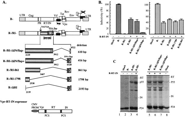
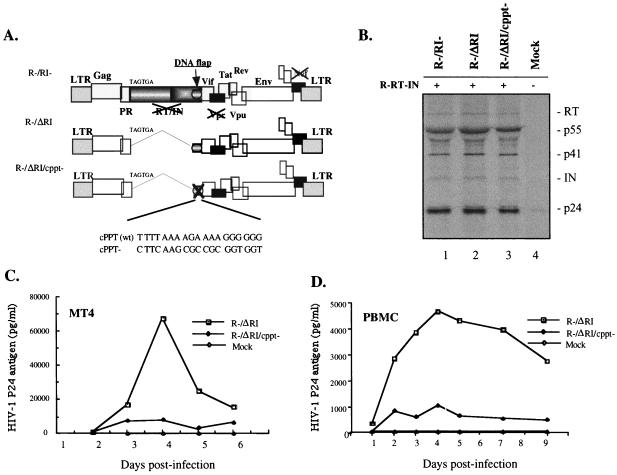
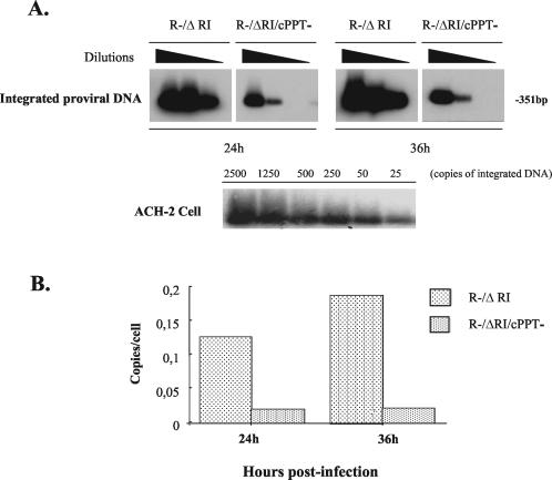
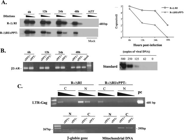
In this study, reverse transcriptase (RT)- and integrase (IN)-defective human immunodeficiency virus type 1 (HIV-1) was transcomplemented with Vpr-RT-IN fusion proteins to delineate pol sequences important for HIV-1 replication. Our results reveal that a 194-bp sequence encompassing the 3'end of the IN gene and containing the central DNA flap is necessary and sufficient for efficient HIV-1 single-cycle replication in dividing and nondividing cells. Furthermore, we show that the central DNA flap enhances HIV-1 single-round replication by five- to sevenfold, primarily by facilitating nuclear import of proviral DNA. In agreement with previous reports, our data support a functional role of the central DNA flap during the early stages of HIV-1 infection.


Figures

FIG. 1.
FIG. 1.
Effect of the HIV-1 IN and RT gene sequences on the infectivity of RT/IN-transcomplemented virus. (A) Schematic structure of HIV-1 proviruses carrying a mutation and/or deletions in pol and of the plasmid encoding the Vpr-RT-IN fusion protein. Provirus R/RI was constructed by replacing the first two amino acids of RT with two premature stop codons (TGA TAG) in HxBruR(R) provirus. In R/RI/ΔIN/flap provirus, a 610-bp fragment of the IN gene sequence (including cPPT/CTS) was deleted. The R/RI/ΔIN/flap+, R/RI-861, R/RI-1798, and R/ΔRI proviruses harbor different deletions within the RT and/or IN gene sequences but contain the 194-bp sequence in the 3′-end region of IN, which harbors the cPPT/CTS cis-acting elements. PR, protease. (B) The infectivity of trans-complemented virus produced in 293T cells was evaluated by MAGI assay. Equal amounts (15 ng of p24gag antigen) of the different viruses were used to infect HeLa-CD4-β-Gal cells, and the number of infected cells was monitored by X-Gal (5-bromo-4-chloro-3-indolyl-β-d-galactopyranoside) staining. The infectivity (% infectivity) of each virus stock was calculated as the ratio of the number of β-Gal-positive cells relative to the number of β-Gal-positive cells obtained with the wt virus (R). The number of β-Gal-positive cells detected with the R virus ranged between 330 to 410 and was set at 100%. The results are representative of three independent experiments. (C) To evaluate Vpr-mediated transincorporation of RT and IN in viral particles, radiolabeled viruses were isolated from cell supernatants, lysed, immunoprecipitated with anti-HIV antibodies, and analyzed by sodium dodecyl sulfate-polyacrylamide gel electrophoresis (12.5% acrylamide). LTR, long terminal repeat.
FIG. 2.
FIG. 2.
The central DNA flap contributes to efficient HIV-1 single-cycle replication. (A) Schematic structure of HIV-1 provirus with the RT/IN gene deleted (R/ΔRI) and the cPPT mutant (R/ΔRI/cPPT). The cPPT element was inactivated by introduction of ten nucleotide substitution mutations, as indicated. (B) To evaluate RT and IN transincorporation, [35S]-methionine-radiolabeled viruses were collected from transfected 293T cells, lysed, and analyzed by immunoprecipitation by using anti-HIV antibodies. To test the replication potential of each virus stock, CD4+ MT4 T cells (C) or PHA-stimulated human PBMCs (D) were infected with equal amounts of R/ΔRI or R/ΔRI/cPPT viruses. At different time intervals after infection, viral production was monitored in the supernatants by measurement of HIV-1 p24gag antigen by using a p24 ELISA assay. The results are representative of two independent experiments.
FIG. 3.
FIG. 3.
Effect of the central DNA flap on HIV-1 proviral DNA integration in h-PBMC. (A) h-PBMCs were infected with R/ΔRI or R/ΔRI/cPPT virus (125 ng of p24/106 cells). At 24- and 36-h p.i., the cells were lysed, and serially diluted cell lysates were analyzed by two-step Alu-PCR and Southern blotting for specific detection of integrated proviral DNA from infected PBMCs (upper panel) or from ACH-2 cells as the quantitative control (lower panel). (B) Quantitative analysis of integrated proviral DNA in single-cycle infection. The bands in panel A were quantified by laser densitometry, and the number of integrated proviral DNA copies per cell was determined by using the PCR-generated standard curve derived from ACH-2 cells.
FIG. 4.
FIG. 4.
Effect of the central DNA flap on HIV-1 cDNA nuclear import. (A) h-PBMCs were infected with transcomplemented R/ΔRI (cPPT+) and R/ΔRI/cPPT (cPPT) viruses (125 ng of p24/106 cells) for 2 h. As the negative control, 3′-azido-3′-deoxythymidine (AZT; 10 μM)-pretreated PBMCs were infected with the same amounts of transcomplemented R/ΔRI virus. At each indicated time point, serial dilutions of extracted total DNA were analyzed for late-reverse-transcription products by PCR by using long terminal repeat (LTR)-Gag primers and Southern blotting. HIV-1 late-reverse-transcription products detected in the left panel were quantified by laser densitometry. The diagram at the right shows the number of HIV-1 cDNA copies per cell as determined by using the PCR-generated standard curve (B, right panel). These results are representative of those obtained in two independent experiments. Serially diluted R-/ΔRI plasmid DNA was used as a standard for DNA copy quantification (right panel). To evaluate cellular DNA levels in each sample, the cellular β2-AR gene was amplified by PCR and visualized by ethidium bromide staining (left panel). (C) At 24-h p.i., 2 × 10⁶ infected h-PBMCs were fractionated into cytoplasmic and nuclear fractions as described previously (36). The amounts of viral DNA in the cytoplasmic and nuclear fractions were evaluated by PCR by using HIV-1 LTR-Gag primers and were visualized by ethidium bromide staining. The R/ΔRI plasmid DNA was used as a PCR-positive control (pc) (upper panel). In parallel, the purity and DNA content of each subcellular fraction were evaluated by PCR detection of the human globin gene and mitochondrial DNA and were visualized by ethidium bromide staining (lower panel). N, nuclear fraction; C, cytoplasmic fraction.

References

    1. Ansari-Lari, M. A., L. A. Donehower, and R. A. Gibbs. 1995. Analysis of human immunodeficiency virus type 1 integrase mutants. Virology 211:332-335. - PubMed
    1. Ansari-Lari, M. A., and R. A. Gibbs. 1996. Expression of human immunodeficiency virus type 1 reverse transcriptase in trans during virion release and after infection. J. Virol. 70:3870-3876. - PMC - PubMed
    1. Bouyac-Bertoia, M., J. D. Dvorin, R. A. Fouchier, Y. Jenkins, B. E. Meyer, L. I. Wu, M. Emerman, and M. H. Malim. 2001. HIV-1 infection requires a functional integrase NLS. Mol. Cell 7:1025-1035. - PubMed
    1. Bukovsky, A., and H. Gottlinger. 1996. Lack of integrase can markedly affect human immunodeficiency virus type 1 particle production in the presence of an active viral protease. J. Virol. 70:6820-6825. - PMC - PubMed
    1. Bukrinsky, M., and O. K. Haffar. 1998. HIV-1 nuclear import: matrix protein is back on center stage, this time together with Vpr. Mol. Med. 4:138-143. - PMC - PubMed

Publication types

MeSH terms

LinkOut - more resources