. 2004 Mar 23;101(12):4065-70.
doi: 10.1073/pnas.0400358101. Epub 2004 Feb 27.

Elastic coupling of integral membrane protein stability to lipid bilayer forces

Affiliations

Elastic coupling of integral membrane protein stability to lipid bilayer forces

Heedeok Hong et al. Proc Natl Acad Sci U S A. .

Abstract

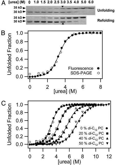
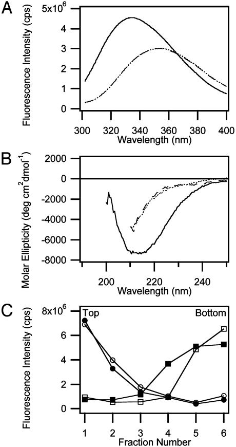
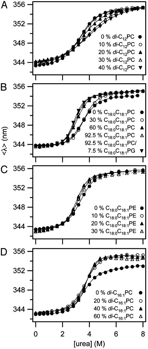
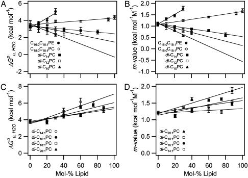
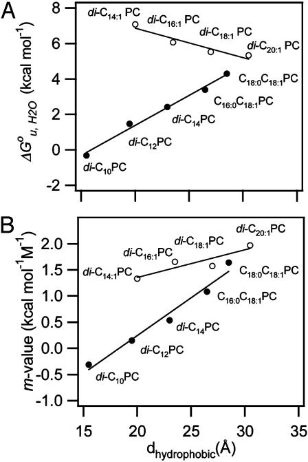
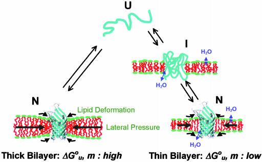
It has been traditionally difficult to measure the thermodynamic stability of membrane proteins because fully reversible protocols for complete folding these proteins were not available. Knowledge of the thermodynamic stability of membrane proteins is desirable not only from a fundamental theoretical standpoint, but is also of enormous practical interest for the rational design of membrane proteins and for optimizing conditions for their structure determination by crystallography or NMR. Here, we describe the design of a fully reversible system to study equilibrium folding of the outer membrane protein A from Escherichia coli in lipid bilayers. Folding is shown to be two-state under appropriate conditions permitting data analysis with a classical folding model developed for soluble proteins. The resulting free energy and m value, i.e., a measure of cooperativity, of unfolding are DeltaG(u,H2O)(o)=3.4 kcal/mol and m = 1.1 kcal/mol M(-1), respectively, in a reference bilayer composed of palmitoyl-oleoyl-phosphatidylcholine (C(16:0)C(18:1)PC) and palmitoyloleoyl-phosphatidylglycerol (C(16:0)C(18:1)PG). These values are strong functions of the lipid bilayer environment. By systematic variation of lipid headgroup and chain composition, we show that elastic bilayer forces such as curvature stress and hydrophobic mismatch modulate the free energy and cooperativity of folding of this and perhaps many other membrane proteins.


Figures

Fig. 1.
Fig. 1.
Reversibility and two-state behavior of OmpA folding in lipid bilayer membranes composed of 92.5% C16:0C18:1PC and 7.5% C16:0C18:1PG at pH 10 and 37.5°C. (A) Comparison of equilibrium unfolding and refolding of OmpA monitored by SDS/PAGE without sample boiling. The 30-kDa form represents the native state, and the 35-kDa form represents denatured states. Transition midpoints are indicated with arrows. (B) Comparison of the urea-induced equilibrium unfolding of OmpA in lipid bilayers monitored by SDS/PAGE (open circles) and Trp fluorescence (filled circles). (C) Same as B, but with increasing mol fractions of the short-chain lipid diC12PC. For clarity, successive curves are each shifted on the abscissa by +1 M urea.
Fig. 2.
Fig. 2.
Characterization of denatured states of OmpA. (A) Fluorescence spectra of native membrane (C16:0C18:1PC/PG, 92:5:7.5)-inserted OmpA (solid line), denatured OmpA obtained by treating native membrane-inserted OmpA with 8 M urea (dashed line), and denatured OmpA in the absence of lipids (dotted line). The emission maxima of the native and denatured states were 333.6 nm and 353.6 nm, respectively. (B) Far-UV CD spectra of native membrane-inserted OmpA (solid line), denatured OmpA obtained by treating native membrane-inserted OmpA with 6 M urea (dashed line), and denatured OmpA in the absence of lipids (dotted line). (C) Airfuge flotation assay to measure degree of membrane-association of denatured OmpA in 6 M urea. NBD-labeled vesicles in the absence (open circles) or presence (filled circles) of denatured OmpA float to the top, and denatured OmpA in the absence (open squares) or presence (filled squares) of vesicles remains at the bottom, indicating separation of denatured OmpA from the vesicles.
Fig. 3.
Fig. 3.
Representative unfolding curves measured by Trp fluorescence with increasing mol fractions of a short-chain saturated PC (A), a long-chain mono-unsaturated PC (B), a long-chain mono-unsaturated PE (C), and a double-unsaturated PC (D). The C16:0C18:1PG fraction was kept fixed at 7.5% in AC and at 12.5% in D. The remainder of the bilayer was filled with C16:0C18:1PC. All experiments were carried out at 37.5°C.
Fig. 4.
Fig. 4.
Dependence of formula image and m-value of OmpA unfolding on lipid composition. Effect of saturated and mono-unsaturated PCs and PEs on formula image (A) and m (B). Effect of cis-double-unsaturated PCs on formula image (C) and m (D). All experiments were carried out at 37.5°C.
Fig. 5.
Fig. 5.
Dependence of formula image (A) and m-value (B) on the hydrophobic thickness of PC bilayers with saturated and mono-unsaturated acyl chains (filled circles) and cis-double-unsaturated acyl chains (open circles).
Fig. 6.
Fig. 6.
Cartoon depicting structures and bilayer forces acting on OmpA folding/unfolding under equilibrium conditions. When folding into most bilayers (left path), the process is two-state. The large black arrows indicate lateral bilayer pressure imparted on the lipid/protein interface in the hydrophobic core (red) of bilayers composed of lipids with negative intrinsic spontaneous curvature. Increasing this pressure increases the thermodynamic stability of the protein. The small black arrows indicate lipid deformation forces caused by hydrophobic mismatch between the protein and unstressed bilayers. These forces decrease the thermodynamic stability of the protein. When folding into thin bilayers (right path), the process is multistate, i.e., at least one equilibrium intermediate occurs. Water molecules penetrate more easily into the hydrophobic core (blue arrows) of more flexible and more dynamic thin bilayers, stabilizing equilibrium intermediates, decreasing the m- and formula image values until, in very thin bilayers composed of saturated lipids, complete unfolding (second step) can no longer be observed under any of our experimental conditions.

Comment in

  • Membrane proteins: a new method enters the fold.
    Bowie JU. Bowie JU. Proc Natl Acad Sci U S A. 2004 Mar 23;101(12):3995-6. doi: 10.1073/pnas.0400671101. Epub 2004 Mar 15. Proc Natl Acad Sci U S A. 2004. PMID: 15024105 Free PMC article. No abstract available.

References

    1. Martinac, B., Adler, J. & Kung, C. (1990) Nature 348, 261–263. - PubMed
    1. Perozo, E., Cortes, D. M., Sompornpisut, P., Kloda, A. & Martinac, B. (2002) Nature 418, 942–948. - PubMed
    1. Keller, S. L., Berzrukov, S. M., Gruner, S. M., Tate, M. W., Vodyanoy, I. & Parsegian, V. A. (1993) Biophys. J. 65, 23–27. - PMC - PubMed
    1. Brown, M. F. (1994) Chem. Phys. Lipids 73, 159–180. - PubMed
    1. Navarro, J., Toivio-Kinnucan, M. & Racker, E. (1984) Biochemistry 23, 130–135. - PubMed

Publication types

MeSH terms

Substances