

Inhibition of neuronal phenotype by PTEN in PC12 cells
- PMID: 14990793
- PMCID: PMC373513
- DOI: 10.1073/pnas.0308289101
Inhibition of neuronal phenotype by PTEN in PC12 cells
Abstract
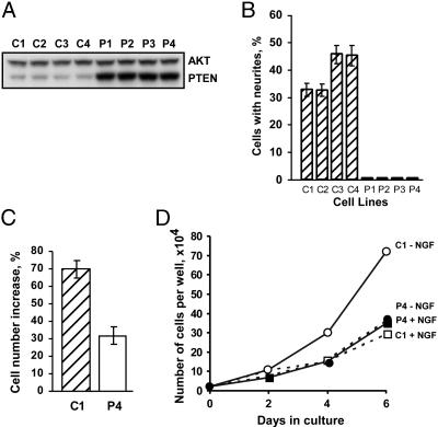
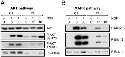
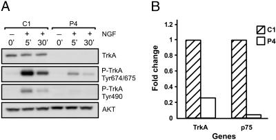
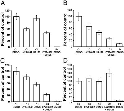
The mechanisms of neuronal differentiation in PC12 cells are still not completely understood. Here, we report that the tumor suppressor PTEN has a profound effect on differentiation by affecting several pathways involved in nerve growth factor (NGF) signaling. When overexpressed in PC12 cells, PTEN (phosphatase and tensin homologue deleted on chromosome ten) blocked neurite outgrowth induced by NGF. In addition, these cells failed to demonstrate the transient mitogenic response to NGF, as well as subsequent growth arrest. Consistent with these observations was a finding that PTEN significantly inhibits NGF-mediated activation of the members of mitogen-activated protein kinase kinase (MEK)/mitogen-activated protein kinase (MAPK) and phosphoinositide 3-kinase (PI3K)/AKT signaling pathways, crucial for these processes. While exploring possible mechanisms of PTEN effects on NGF signaling, we discovered a significant down-regulation of both high-affinity (TrkA) and low-affinity (p75) NGF receptors in PTEN-overexpressing clones. Subsequent microarray analysis of several independent clonal isolates revealed a myriad of neuronal genes to be affected by PTEN. All of these changes were validated by quantitative PCR. Of particular interest were the genes for the key enzymes of the dopamine synthesis pathway, receptors for different neurotransmitters, and neuron-specific cytoskeleton proteins, among others. Some, but not all effects could be reproduced by pharmacological inhibitors of PI3K and/or MAPK, suggesting that PTEN may influence some genes by mechanisms independent of these signaling pathways. Our findings may shed new light on the role of this tumor suppressor during normal brain development and suggest a previously uncharacterized mechanism of PTEN action in neuron-like cells.
Figures
References
-
- Li, J., Yen, C., Liaw, D., Podsypanina, K., Bose, S., Wang, S. I., Puc, J., Miliaresis, C., Rodgers, L., McCombie, R., et al. (1997) Science 275, 1943-1947. - PubMed
-
- Maehama, T. & Dixon, J. E. (1998) J. Biol. Chem. 273, 13375-13378. - PubMed
-
- Stambolic, V., Suzuki, A., de la Pompa, J. L., Brothers, G. M., Mirtsos, C., Sasaki, T., Ruland, J., Penninger, J. M., Siderovski, D. P. & Mak, T. W. (1998) Cell 95, 29-39. - PubMed
Publication types
MeSH terms
Substances
LinkOut - more resources
Full Text Sources
Other Literature Sources
Molecular Biology Databases
Research Materials
