. 2004 Mar 9;101(10):3557-62.
doi: 10.1073/pnas.0306431101. Epub 2004 Feb 27.

Human CD34+ cells differentiate into microglia and express recombinant therapeutic protein

Affiliations

Human CD34+ cells differentiate into microglia and express recombinant therapeutic protein

Muriel Asheuer et al. Proc Natl Acad Sci U S A. .

Abstract

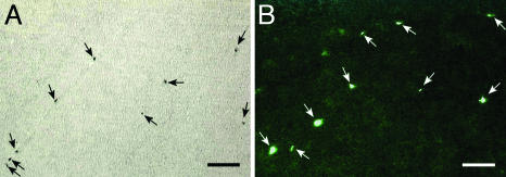
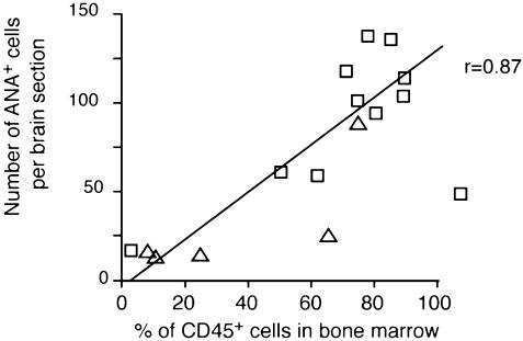
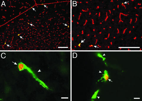
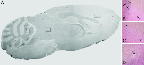
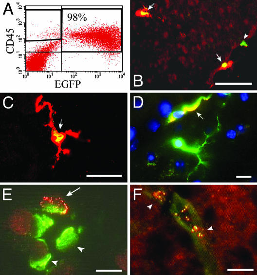
In rodents, bone marrow-derived cells enter the brain during adult life. Allogeneic bone marrow transplantation is used to treat genetic CNS diseases, but the fate of human bone marrow and CD34(+) cells within the brain remains to be elucidated. The present study demonstrates that cells derived from human CD34(+) cells, isolated from either cord blood or peripheral blood, migrate into the brain after infusion into nonobese diabetic/severe combined immunodeficient mice. Both types of CD34(+)-derived cells differentiate into perivascular and ramified microglia. The lentiviral transfer of genes into CD34(+) cells before infusion does not modify the differentiation of human CD34(+) cells into microglia, allowing new transgenic proteins to be expressed in these cells. The transplantation of CD34(+) cells could thus be used for the treatment of CNS diseases.


Figures

Fig. 1.
Fig. 1.
Engraftment of human CD34+ cells in the brain of NOD/SCID mice. The human origin of the engrafted cells was established by using in situ hybridization with an Alu probe (A) or by immunohistochemistry by using the ANA marker (B). (Bars = 100 μm.)
Fig. 2.
Fig. 2.
Cerebral homing of human CD34+-derived cells is correlated to bone marrow engraftment of UCB (□) or MPB (▵) CD34+ cells. Human cell engraftment was assessed by scoring the number of human CD45+ cells in the bone marrow of NOD/SCID mice, 18 weeks after transplantation. Brain engraftment was assessed by scoring the number of ANA-positive cells in the brain of recipient mice at the same time. In two animals, the brain engraftment was limited despite high levels of bone marrow engraftment.
Fig. 3.
Fig. 3.
Differentiation of human CD34+-derived cells in the brain of NOD/SCID mice. (A and B) Double-labeling of brain sections identifies cells coexpressing the microglial marker RCA-1 (in red, Cy3) and the human ANA marker (in green, FITC). Yellow staining (arrows) indicates colocalization of both markers in the same cell. (C and D) Double-labeling of brain sections identifies cells coexpressing the microglial marker Iba1 (in green, FITC, arrowheads) and the ANA marker (in red, Cy3, arrows) in their nuclei.
Fig. 4.
Fig. 4.
Distribution of human CD34+-derived cells in the brain of NOD/SCID mice. (A) Human-engrafted cells, detected by in situ hybridization with Alu probe, were widely distributed throughout the brain. One hundred forty-one human cells are detected on this brain section. This section is representative of the brain of mice transplanted with human UCB CD34+ cells (≥80% of engraftment) (see Table 1). (B-D) Large magnification. Engrafted cells were located at perivascular sites as well as in the brain parenchyma (B, cerebellum; C, colliculus; D, cortex). Perivascular (B and D, arrows) and intraparenchymatous (B and C, arrowheads) engrafted cells were identified on the basis of morphology and proximity to or absence of nearby blood vessels.
Fig. 5.
Fig. 5.
Genetically modified human CD34+-derived microglial cells express marker or therapeutic genes in the brains of NOD/SCID mice. (A) Dot-plot analysis showing the percentage of human CD45+ cells expressing EGFP in mouse bone marrow. (B and C) EGFP-transduced UCB CD34+-derived cells in mouse brain. Nonramified (B) and ramified (C) cells express EGFP (in red, Cy3) and the human ANA marker (green, FITC) yielding a yellow staining in their nuclei (arrows). One ANA-positive cell (B, arrowhead) does not express EGFP. (D) Human UCB CD34+-derived cells differentiate into microglia expressing EGFP. Colocalization of EGFP (in red, Cy3) and Iba1 (in green, FITC) results in yellow staining (arrow). (E) CD68+ macrophages (in green, FITC, arrowheads) expressing recombinant ALD protein (punctate staining in red, Cy3, arrow) are present in the bone marrow of transplanted mice. (F) MPB CD34+ cells from ALD patient engrafted in mouse brain express recombinant ALD protein (punctate staining in red, Cy3, arrows) and the microglial RCA-1 lectin (in green, FITC). [Bars = 100 μm (B), 25 μm (C), and 10 μm (D-F).]

Similar articles

Cited by

References

    1. Lawson, L. J., Perry, V. H., Dri, P. & Gordon, S. (1990) Neuroscience 39, 151-170. - PubMed
    1. Perry, V. H. & Gordon, S. (1991) Int. Rev. Cytol. 125, 203-244. - PubMed
    1. Eglitis, M. A. & Mezey, E. (1997) Proc. Natl. Acad. Sci. USA 94, 4080-4085. - PMC - PubMed
    1. Brazelton, T. R., Rossi, F. M., Keshet, G. I. & Blau, H. M. (2000) Science 290, 1775-1779. - PubMed
    1. Mezey, E., Chandross, K. J., Harta, G., Maki, R. A. & McKercher, S. R. (2000) Science 290, 1779-1782. - PubMed

Publication types

MeSH terms