. 2004 Mar;113(5):694-700.
doi: 10.1172/JCI19210.

Local expression of B7-H1 promotes organ-specific autoimmunity and transplant rejection

Affiliations

Local expression of B7-H1 promotes organ-specific autoimmunity and transplant rejection

Sumit K Subudhi et al. J Clin Invest. 2004 Mar.

Abstract

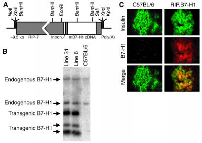
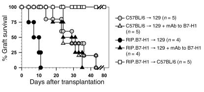
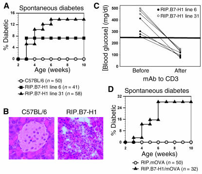
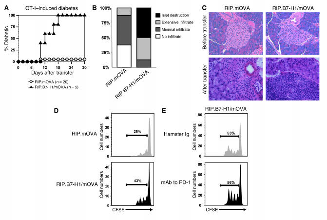
A number of studies have suggested B7-H1, a B7 family member, inhibits T cell responses. Therefore, its expression on nonlymphoid tissues has been proposed to prevent T cell-mediated tissue destruction. To test this hypothesis, we generated transgenic mice that expressed B7-H1 on pancreatic islet beta cells. Surprisingly, we observed accelerated rejection of transplanted allogeneic B7-H1-expressing islet beta cells. Furthermore, transgenic B7-H1 expression broke immune tolerance, as some of the mice spontaneously developed T cell-dependent autoimmune diabetes. In addition, B7-H1 expression increased CD8+ T cell proliferation and promoted autoimmunity induction in a T cell adoptive transfer model of diabetes. Consistent with these findings, B7-H1.Ig fusion protein augmented naive T cell priming both in vitro and in vivo. Our results demonstrate that B7-H1 can provide positive costimulation for naive T cells to promote allograft rejection and autoimmune disease pathogenesis.


Figures

Figure 1
Figure 1
Generation of RIP.B7-H1 transgenic mice. (A) A schematic of the RIP–murine B7-H1 hybrid gene. (B) Southern blot analysis of mouse genomic DNA digested with BamHI and hybridized with a full-length murine B7-H1 probe. (C) Immunofluorescence staining of insulin (green) and murine B7-H1 (red) in pancreata (original magnification, ×40) from C57BL/6 and RIP.B7-H1 mice.
Figure 2
Figure 2
Accelerated rejection of B7-H1–expressing pancreatic β cells in an islet allograft setting. Isolated islet β cells from C57BL/6 and RIP.B7-H1 (line 31) mice were transplanted into 129 recipient mice (C57BL/6 → 129 and RIP.B7-H1 → 129, respectively), and graft survival was determined. As indicated, some of the mice were treated with mAb to B7-H1. As an isograft control, islets from RIP.B7-H1 mice were transplanted into syngeneic C57BL/6 mice (RIP.B7-H1 → C57BL/6). The statistical significance between groups was determined using the Kaplan-Meier log-rank test method.
Figure 3
Figure 3
RIP.B7-H1 mice develop T cell–mediated spontaneous diabetes. (A) Incidence of spontaneous diabetes was evaluated in RIP.B7-H1 mice. (B) H&E staining (original magnification, ×40) of pancreata from C57BL/6 and diabetic RIP.B7-H1 mice. (C) Treatment of diabetic RIP.B7-H1 mice with mAb to CD3. As indicated in the graph, blood glucose levels were detected immediately before treatment onset and 4 days after treatment termination. (D) Incidence of spontaneous diabetes was evaluated in RIP.B7-H1/mOVA mice.
Figure 4
Figure 4
B7-H1 expression in the pancreatic islets promotes CD8+ T cell priming and autoimmune diabetes induction. (A) OT-I T cells (2 × 10⁶) were adoptively transferred into RIP.mOVA and RIP.B7-H1/mOVA mice. Incidence of diabetes was evaluated at daily intervals. (B) Pancreata were stained with H&E, and individual islets from three mice per group were assigned scores for insulitis at the time of diabetes onset (RIP.B7-H1/mOVA mice) or day 22 after transfer for those mice that did not develop diabetes. (C) H&E staining of pancreata from RIP.mOVA and RIP.B7-H1/mOVA mice before and after transfer of 2 × 10⁶ OT-I T cells. Original magnifications: top panels, ×20; bottom panels, ×40. (D) CFSE-labeled OT-I T cells (2 × 10⁶) were adoptively transferred into RIP.mOVA and RIP.B7-H1/mOVA mice. Then, 42 hours later, the pancreatic draining lymph node cells were analyzed by flow cytometry. The numbers indicate the percentage of dividing OT-I T cells. (E) CFSE-labeled OT-I T cells (2 × 10⁶) were adoptively transferred into RIP.B7-H1/mOVA mice. At the time of transfer, the mice were treated with 100 μg hamster IgG (top panel) or mAb to mouse PD-1 (bottom panel). Then, 42 hours later, the pancreatic draining lymph node cells were analyzed by flow cytometry. The numbers indicate the percentage of dividing OT-I T cells. Each of the experiments was repeated at least three times.
Figure 5
Figure 5
B7-H1.Ig provides T cell costimulation both in vitro and in vivo. (A) CFSE-labeled OT-I T cells (2 × 10⁶) were adoptively transferred into RIP.mOVA mice treated with 20 μg control Ig or B7-H1.Ig intraperitoneally at the time of transfer. Then, 42 hours later, the pancreatic draining lymph node cells were analyzed by flow cytometry. The numbers indicate the percentage of dividing OT-I T cells. (B–D) Purified CD3+ T cells (B), CD4+ T cells (C), and CD8+ T cells (D) from C57BL/6 mice were stimulated with bead-bound anti-CD3/control Ig or anti-CD3/B7-H1.Ig for 96 hours. The statistical significance between groups was determined using Student’s t-test: *P < 0.05; **P < 0.01.

References

    1. Dong H, Zhu G, Tamada K, Chen L. B7-H1, a third member of the B7 family, co-stimulates T-cell proliferation and interleukin-10 secretion. Nat. Med. 1999;5:1365–1369. - PubMed
    1. Dong H, et al. Tumor-associated B7-H1 promotes T-cell apoptosis: a potential mechanism of immune evasion. Nat. Med. 2002;8:793–800. - PubMed
    1. Tamura H, et al. B7-H1 costimulation preferentially enhances CD28-independent T-helper cell function. Blood. 2001;97:1809–1816. - PubMed
    1. Moore KW, de Waal Malefyt R, Coffman RL, O’Garra A. Interleukin-10 and the interleukin-10 receptor. Annu. Rev. Immunol. 2001;19:683–765. - PubMed
    1. Freeman GJ, et al. Engagement of the PD-1 immunoinhibitory receptor by a novel B7 family member leads to negative regulation of lymphocyte activation. J. Exp. Med. 2000;192:1027–1034. - PMC - PubMed

Publication types

MeSH terms